TSH and Free T4 Patterns: Solve Thyroid Lab Questions
Compare primary, central, and subclinical thyroid lab patterns. Learn when normal TSH misleads, when to check T3, and how to solve a worked USMLE vignette.
High TSH with low free T4 indicates primary hypothyroidism; low free T4 with a TSH that is not appropriately elevated suggests central hypothyroidism. An abnormal TSH with normal free T4 may indicate subclinical disease, but a low TSH also requires T3 assessment before calling the pattern subclinical hyperthyroidism. The essential distinction is whether the pituitary response fits the thyroid hormone level—not simply whether each number falls inside its reference interval. These relationships underpin NIDDK’s explanation of thyroid testing and the central hypothyroidism diagnostic guidelines.
Why TSH and free T4 usually move in opposite directions
The hypothalamic–pituitary–thyroid axis is a feedback circuit. Hypothalamic thyrotropin-releasing hormone stimulates pituitary TSH secretion, and TSH stimulates thyroid hormone production. Thyroid hormones feed back to reduce upstream stimulation. With an intact axis, falling free T4 therefore increases TSH, while rising free T4 suppresses it. The European Thyroid Association’s Graves hyperthyroidism guideline describes this inverse relationship: relatively small free T4 changes can produce much larger TSH changes.
This sensitivity explains why TSH can become abnormal before free T4 leaves the population reference interval. It also explains why “normal TSH” is not universally reassuring: that interpretation assumes the feedback machinery works. In suspected pituitary disease, both TSH and free T4 should be measured, as recommended in the NICE thyroid disease guideline.
Use two separate questions:
- **Free T4:** Is circulating unbound thyroxine low, normal, or high?
- **TSH:** Is the pituitary responding appropriately to that concentration?
The second question localizes the problem. For example, a TSH of 2.0 mIU/L may be within the laboratory interval, but it is an inadequate response to a clearly low free T4. A clinical investigation of TSH-only testing illustrates how this approach can miss secondary hypothyroidism.
TSH and free T4 patterns: primary, central, and subclinical
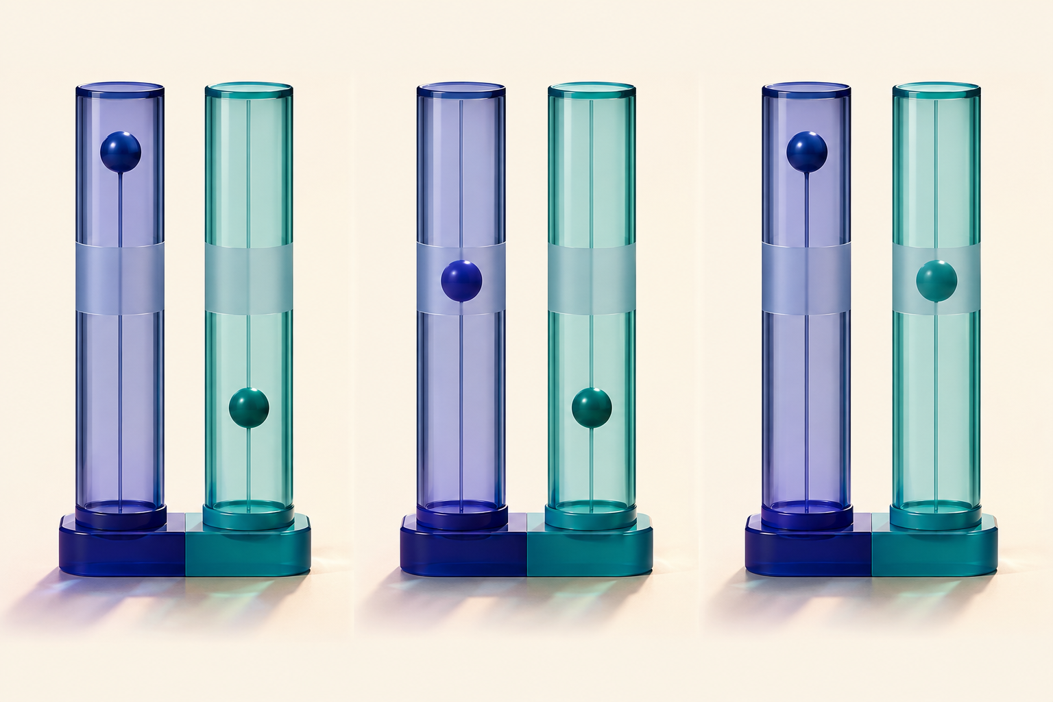
Interpret arrows relative to the laboratory’s reference intervals. The table assumes no recent treatment change, major acute illness, or assay interference; those situations can disrupt the expected relationship.
| Pattern | TSH | Free T4 | Interpretation or next distinction | |---|---|---|---| | Overt primary hypothyroidism | High | Low | Thyroid output is inadequate despite increased pituitary stimulation | | Central hypothyroidism | Low, normal, or occasionally mildly high | Low | TSH stimulation is inadequate for the degree of hormone deficiency | | Subclinical primary hypothyroidism | High | Normal | Abnormal feedback signal without low circulating free T4 | | Overt primary hyperthyroidism | Suppressed | High, or normal if T3 alone is elevated | Check T3 when free T4 is normal | | Subclinical hyperthyroidism | Low | Normal | T3 must also be normal; establish persistence | | Elevated hormones with nonsuppressed TSH | Normal or high | High | Exclude interference; consider a TSH-secreting tumor or thyroid hormone resistance |
The primary patterns are summarized by NIDDK. The central and subclinical distinctions follow the central hypothyroidism, subclinical hypothyroidism, and subclinical hyperthyroidism guidelines; the discordant high-hormone pattern is addressed in the TSH-secreting pituitary tumor guideline.
Primary disease: the pituitary response is appropriate
In primary hypothyroidism, the thyroid cannot produce enough hormone. Low free T4 reduces negative feedback, so TSH rises. In primary hyperthyroidism, excess thyroid hormone suppresses pituitary TSH. Thus, the pituitary is responding correctly even though thyroid output is abnormal. These are the opposing patterns described in NIDDK’s thyroid test guidance.
The pair identifies functional status, not the exact cause. For example, autoimmune testing can help clarify etiology after thyroid dysfunction is established. Do not confuse “this patient is hypothyroid” with “this patient has a proven autoimmune cause.” NIDDK distinguishes thyroid hormone testing from thyroid antibody testing.
Central disease: “normal” TSH can be abnormal feedback
Central hypothyroidism includes **secondary disease**, arising in the pituitary, and **tertiary disease**, arising in the hypothalamus. Both can produce low free T4 without the expected TSH elevation. Basal TSH and free T4 generally cannot distinguish these two anatomical levels by themselves. Occasionally, measured TSH is mildly elevated but has reduced biological activity; that does not restore an appropriate thyroid response. These features are described in the European Thyroid Association central hypothyroidism guideline.
The key phrase is **inappropriately normal TSH**. The reference interval describes a population; it does not establish whether the pituitary response is appropriate for this patient’s low free T4. Consequently, a laboratory algorithm that measures free T4 only when TSH is abnormal can miss central disease—a limitation demonstrated in research on routine thyroid profiles.
Subclinical disease: a biochemical category, not a symptom verdict
Subclinical hypothyroidism means elevated TSH with free T4 still within range. It does not mean that symptoms determine the category. An initially elevated TSH should usually be confirmed, because transient abnormalities can occur. The subclinical hypothyroidism guideline recommends repeating TSH and free T4, with thyroid peroxidase antibody assessment, generally after two to three months in appropriate stable patients.
Subclinical hyperthyroidism requires low TSH with **both free T4 and T3 within range**. Low TSH plus normal free T4 alone is incomplete information: elevated T3 changes the diagnosis to overt T3-predominant thyrotoxicosis. The subclinical hyperthyroidism guideline also requires persistence rather than a single transient low TSH result.
When an isolated TSH is insufficient
Suspected pituitary or hypothalamic dysfunction
Measure TSH and free T4 together when central dysfunction is plausible. Prior pituitary surgery, cranial irradiation, or a sellar lesion changes the meaning of a normal TSH. The NICE testing recommendations explicitly distinguish testing for suspected primary dysfunction from testing when secondary dysfunction is suspected.
Suppressed TSH with normal free T4
Add a T3 measurement. The thyroid secretes T4 and T3, and peripheral tissues also generate T3 from T4. A normal T4 concentration does not exclude excess T3; NIDDK specifically identifies T3 testing as useful when hyperthyroidism is suspected despite normal T4.
Severe illness or recent treatment changes
Nonthyroidal illness can lower T3 and, with greater illness severity, lower T4 without an appropriate TSH rise. The pattern can therefore resemble central hypothyroidism. Conversely, TSH can remain suppressed after treatment has lowered elevated thyroid hormones. NICE cautions against interpreting acute-illness abnormalities in isolation, and the Graves guideline describes delayed TSH recovery during treatment.
The practical distinction is clinical context: a stable patient with pituitary disease and a critically ill patient may have similar arrows for different reasons. Confirm the pattern and investigate its setting rather than treating a laboratory label alone.
Binding proteins and assay interference can create misleading results
**Total T4 is not free T4.** Total hormone includes protein-bound and unbound fractions. An isolated increase in binding protein can increase total T4 without producing true hormone excess; free hormone testing helps separate binding changes from dysfunction. NIDDK explains why free T4 is useful when binding proteins alter total hormone measurements.
Pregnancy deserves an additional qualification. Estrogen increases thyroxine-binding globulin, while hCG can stimulate the thyroid. Pregnancy is therefore not simply an isolated binding-protein experiment; results require pregnancy-appropriate interpretation. NIDDK’s pregnancy guidance explains these physiological changes.
**Biotin can create a false hyperthyroid-looking pattern.** On susceptible assays, supplementation can produce falsely low TSH and falsely high thyroid hormone measurements. The effect depends on the assay platform, as demonstrated in a prospective biotin interference study. Discordance between symptoms and laboratory results should prompt review of supplements and laboratory methods before assigning a diagnosis.
Finally, elevated free T4 with nonsuppressed TSH is not the ordinary primary hyperthyroid pattern. After excluding interference and other confounders, consider a TSH-secreting pituitary tumor or thyroid hormone resistance. Resistance is not itself a pituitary tumor; the two conditions share a biochemical pattern but differ mechanistically, as explained in the TSH-secreting tumor guideline.
Worked vignette: low free T4 with a normal TSH
*Original educational vignette:* A 38-year-old woman reports fatigue, constipation, and cold intolerance four months after surgery for a nonfunctioning pituitary macroadenoma. She is clinically stable and takes no thyroid medication. TSH is 1.8 mIU/L (reference 0.4–4.5), and free T4 is 0.5 ng/dL (reference 0.8–1.8). Which diagnosis best explains the pair?
**Answer: central hypothyroidism from pituitary dysfunction.**
The decisive clue is not fatigue; it is **low free T4 without a compensatory TSH increase**, reinforced by the pituitary history. The TSH is numerically normal but physiologically inadequate. This is the diagnostic limitation identified in research comparing TSH-only testing with paired testing.
The nearest distractor, primary hypothyroidism, loses because an intact pituitary should substantially increase TSH. Subclinical hypothyroidism also loses: free T4 is already low, not normal. Severe nonthyroidal illness is less persuasive because the patient is stable and has a direct central risk factor.
The management consequence matters: assess for accompanying central adrenal insufficiency before starting levothyroxine; if cortisol deficiency is present or cannot be excluded, glucocorticoid coverage must precede thyroid replacement to avoid precipitating adrenal crisis. This sequence is specified in the central hypothyroidism guideline.
Once central hypothyroidism is treated, free T4 and clinical assessment—not normalization of TSH—guide replacement. A study comparing central and primary hypothyroidism illustrates the risk of under-replacement in pituitary patients.
Final takeaways
- **High TSH + low free T4:** primary hypothyroidism.
- **Low free T4 + inadequately elevated TSH:** consider central hypothyroidism, while excluding illness and testing confounders.
- **Abnormal TSH + normal free T4:** evaluate for subclinical disease; low TSH also requires T3 assessment.
- **Normal TSH is not a universal rule-out:** pituitary disease, illness, treatment timing, and assay interference can invalidate that shortcut.
For related endocrine physiology, explore the CoreStepPrep Core Concepts library.
Apply the feedback distinction to a fresh vignette: Try a free USMLE question at CoreStepPrep.
Sources and further reading
- www.niddk.nih.gov/health-information/diagnostic-tests/thyroid
- pmc.ncbi.nlm.nih.gov/articles/PMC6198777
- pmc.ncbi.nlm.nih.gov/articles/PMC6140607
- www.ncbi.nlm.nih.gov/books/NBK550859
- pubmed.ncbi.nlm.nih.gov/26600054
- pmc.ncbi.nlm.nih.gov/articles/PMC3923601
- pmc.ncbi.nlm.nih.gov/articles/PMC4637513
- pmc.ncbi.nlm.nih.gov/articles/PMC3821512
- www.niddk.nih.gov/-/media/Files/Endocrine-Diseases/Pregnancy_Thyroid_Disease_508.pdf
- pmc.ncbi.nlm.nih.gov/articles/PMC8420951
- pubmed.ncbi.nlm.nih.gov/21521256