Renal Tubular Acidosis Types 1, 2, and 4
Compare renal tubular acidosis types 1, 2, and 4 by potassium, urine pH, mechanism, associations, diagnostic clues, and treatment for Step 1.
Renal tubular acidosis (RTA) causes a **normal-anion-gap, hyperchloremic metabolic acidosis** because the kidney cannot appropriately reclaim bicarbonate or excrete acid. The fastest distinction is potassium plus urine behavior: **type 1 is hypokalemic with persistently alkaline urine, type 2 is hypokalemic with urine pH that changes as serum bicarbonate falls, and type 4 is hyperkalemic because aldosterone activity and ammonium excretion are reduced.**
Why renal tubular acidosis produces a normal anion gap
The serum anion gap is calculated as:
**Anion gap = Na⁺ − (Cl⁻ + HCO₃⁻)**
In RTA, serum bicarbonate decreases, but chloride rises to preserve electroneutrality. The result is metabolic acidosis without a major increase in unmeasured anions. Contemporary reviews therefore classify RTA as a subset of normal-anion-gap metabolic acidosis caused by disturbances in renal bicarbonate reclamation or net acid excretion (Renal Tubular Acidosis: Core Curriculum 2025).
Before assigning an RTA type, confirm that the patient truly has metabolic acidosis rather than an isolated low bicarbonate value. Then consider major nonrenal causes of normal-anion-gap acidosis, especially diarrhea, before using potassium and urine studies to localize the tubular defect.
Renal tubular acidosis types 1, 2, and 4 compared
| Feature | Type 1: distal RTA | Type 2: proximal RTA | Type 4: hyperkalemic RTA | |---|---|---|---| | Primary defect | Failure of distal H⁺ secretion | Reduced proximal HCO₃⁻ reabsorption | Reduced aldosterone effect and impaired NH₄⁺ excretion | | Main nephron site | α-intercalated cells of collecting duct | Proximal convoluted tubule | Collecting duct, with reduced ammonium availability | | Serum potassium | **Low** | **Low** | **High** | | Urine pH during established acidosis | Usually **>5.5** | Usually **<5.5** after bicarbonate depletion | Usually **<5.5** | | Characteristic clue | Calcium phosphate stones or nephrocalcinosis | Fanconi syndrome findings | Diabetes, kidney disease, or an aldosterone-blocking drug | | Common associations | Sjögren syndrome, other autoimmune disease, amphotericin B, obstruction | Carbonic anhydrase inhibitors, tenofovir, ifosfamide, Fanconi syndrome | Hyporeninemic hypoaldosteronism, ACE inhibitors, ARBs, heparin, potassium-sparing diuretics | | General treatment principle | Alkali, often with potassium replacement | Larger alkali requirement plus treatment of proximal losses | Correct hyperkalemia and the aldosterone-related cause |
These patterns reflect three different physiologic failures rather than three arbitrary collections of laboratory findings. A detailed clinical review similarly distinguishes distal acid-secretion failure, proximal bicarbonate wasting, and hyperkalemic impairment of collecting-duct acid and potassium handling (narrative review of RTA mechanisms and management).
Type 1 distal RTA: the nephron cannot lower urine pH
Type 1 RTA results from impaired H⁺ secretion by α-intercalated cells in the distal nephron. Because this is the final site at which urine is acidified, the kidney cannot appropriately lower urine pH despite systemic metabolic acidosis.
The classic pattern is:
- Normal-anion-gap metabolic acidosis
- **Hypokalemia**
- **Urine pH above approximately 5.5 despite acidosis**
- A low rate of urinary ammonium excretion
Persistently alkaline urine promotes **calcium phosphate precipitation**. Chronic acidosis also increases citrate reabsorption, producing hypocitraturia and removing an important inhibitor of stone formation. The combination explains the association with nephrolithiasis and nephrocalcinosis.
Autoimmune disease is an important association. A patient with Sjögren syndrome, weakness from hypokalemia, normal-anion-gap acidosis, and a urine pH of 6.2 has distal RTA until a better explanation appears. Amphotericin B and urinary tract obstruction are other classic settings.
The decisive concept is not merely that the urine is alkaline. It is that the urine remains inappropriately alkaline **while the blood is acidemic**. Urine pH can be elevated by other conditions, including infection with a urease-producing organism, so the value must be interpreted in its clinical setting.
Type 2 proximal RTA: bicarbonate wasting with preserved distal acidification
In type 2 RTA, the proximal tubule cannot reclaim its normal proportion of filtered bicarbonate. More bicarbonate reaches the distal nephron and is lost in urine, lowering the serum bicarbonate concentration.
Potassium falls because increased distal sodium delivery favors sodium reabsorption coupled to potassium secretion. Volume contraction from urinary solute loss can also activate aldosterone, further increasing potassium loss.
Type 2 RTA may occur as an isolated bicarbonate transport defect, but Step 1 vignettes often place it within **Fanconi syndrome**, a generalized proximal tubular disorder. Look for inappropriate urinary losses of substances normally reclaimed in the proximal tubule:
- Glucose despite a normal serum glucose concentration
- Phosphate, potentially causing hypophosphatemia
- Amino acids
- Uric acid
- Low-molecular-weight proteins
- Bicarbonate
Carbonic anhydrase inhibitors cause proximal bicarbonate wasting. Other acquired associations include tenofovir, ifosfamide, heavy-metal toxicity, and monoclonal light-chain disorders.
The type 2 urine-pH caveat
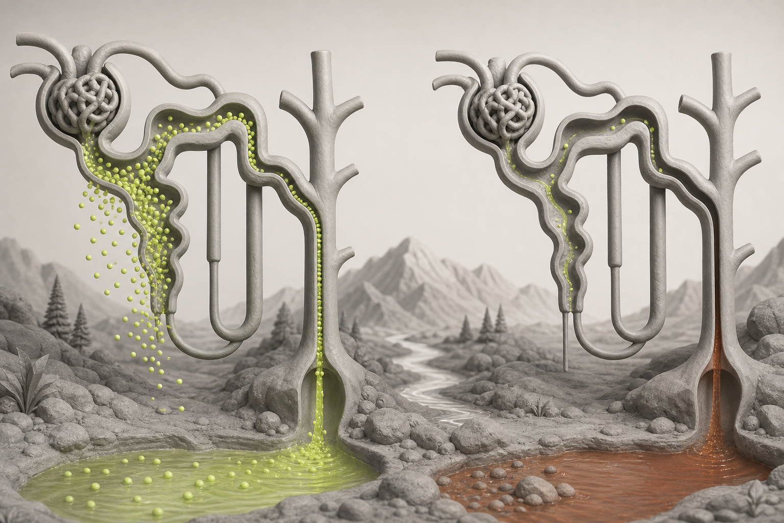
The most important urine-pH trap is assuming that proximal RTA must always produce alkaline urine.
Early in type 2 RTA—or after bicarbonate has been administered—the filtered bicarbonate load exceeds the proximal tubule’s reduced reabsorptive threshold. Bicarbonate spills into urine, so **urine pH rises**.
As bicarbonate loss continues, however, the serum bicarbonate concentration falls below that reduced threshold. The remaining filtered bicarbonate can then be reclaimed, and the intact distal nephron still secretes H⁺. At this new steady state, **urine pH can fall below 5.5 even though the patient still has metabolic acidosis**. This variable urine pH is a defining diagnostic feature of proximal RTA (review of the diagnostic evaluation of RTA).
Think of type 2 RTA as a leaky bicarbonate threshold:
- **Serum bicarbonate above the reduced threshold:** bicarbonaturia and alkaline urine.
- **Serum bicarbonate below the reduced threshold:** little filtered bicarbonate escapes, allowing acidic urine.
- **Bicarbonate treatment:** serum bicarbonate rises again, renewed urinary wasting occurs, and large alkali doses may be required.
Therefore, a urine pH of 5.1 does not exclude proximal RTA. If the vignette includes euglycemic glucosuria, phosphaturia, or another marker of proximal tubular dysfunction, those findings outweigh the simplistic rule that “RTA means alkaline urine.”
Type 4 RTA: hyperkalemia points to reduced aldosterone effect
Type 4 is the major RTA associated with **hyperkalemia**. It results from aldosterone deficiency, impaired renin release, or renal resistance to aldosterone.
Reduced aldosterone activity decreases sodium reabsorption through epithelial sodium channels in principal cells. This reduces the lumen-negative electrical gradient that normally supports potassium secretion. Hyperkalemia also suppresses renal ammonia production, so less NH₃ is available to buffer secreted H⁺ as NH₄⁺. Net acid excretion falls, producing a usually mild normal-anion-gap acidosis.
Common settings include:
- Diabetic kidney disease with hyporeninemic hypoaldosteronism
- ACE inhibitor or ARB therapy
- Potassium-sparing diuretics
- Heparin-associated reduction in aldosterone production
- Adrenal insufficiency
- Tubulointerstitial kidney disease
Unlike classic distal RTA, the distal nephron may retain the ability to lower urine pH. Thus, urine pH is often below 5.5 even though total acid excretion is inadequate. The problem is largely insufficient **ammonium excretion**, not necessarily an inability to create an acidic free urine pH.
A practical diagnostic sequence
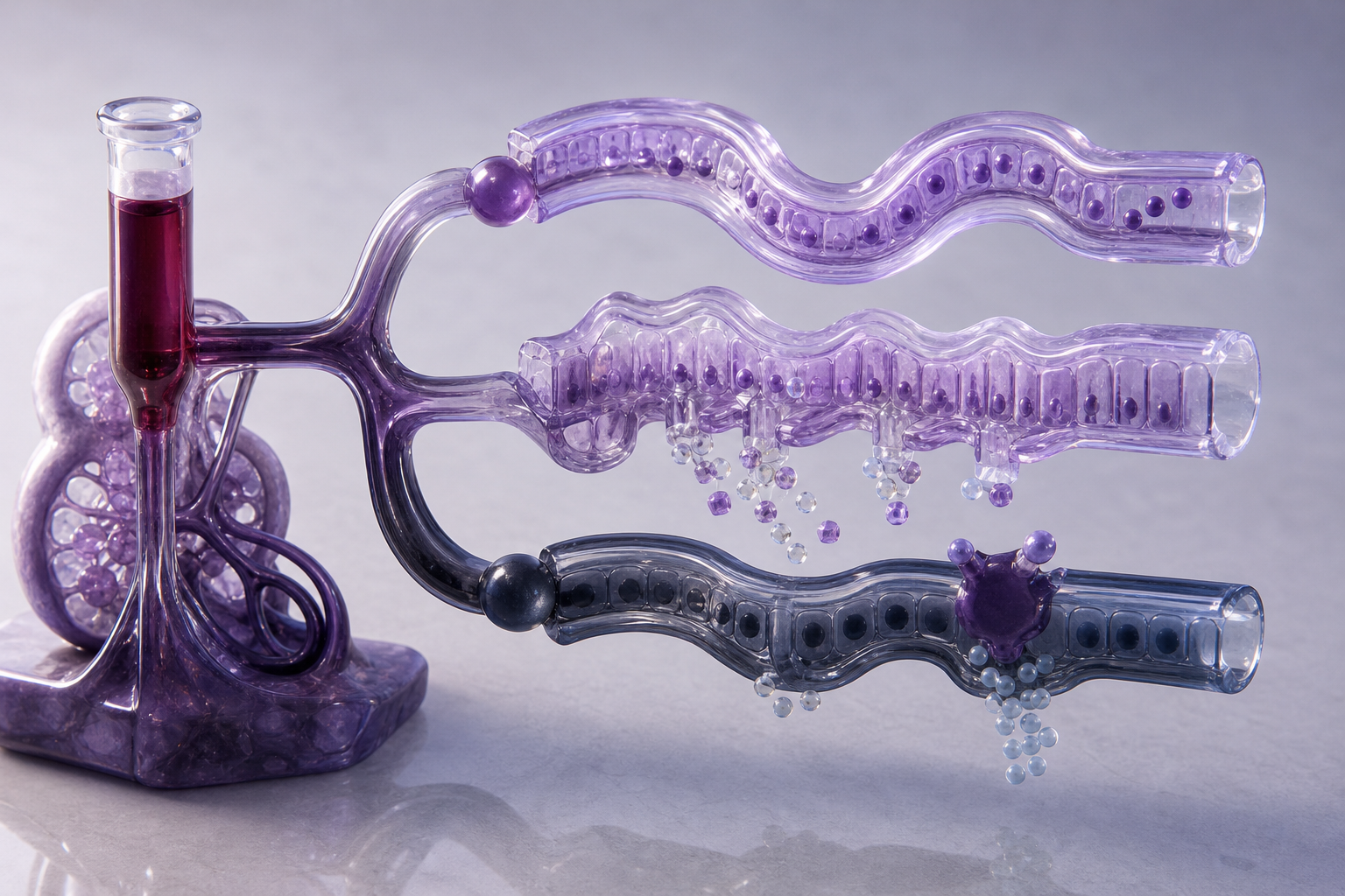
Start with the serum potassium because it immediately divides the differential.
If potassium is low
Consider type 1, type 2, diarrhea, or another source of potassium and bicarbonate loss.
- **Urine pH persistently >5.5 during acidosis:** favors type 1 distal RTA.
- **Urine pH <5.5 with Fanconi findings:** favors type 2 proximal RTA.
- **Evidence of gastrointestinal loss with an appropriate renal ammonium response:** favors diarrhea.
If potassium is high
Consider type 4 RTA, especially when the patient has diabetes, mild-to-moderate kidney dysfunction, adrenal disease, or medication-related interference with the renin-angiotensin-aldosterone system.
The **urine anion gap** can estimate the ammonium response:
**Urine anion gap = urine Na⁺ + urine K⁺ − urine Cl⁻**
NH₄⁺ is generally excreted with chloride. Therefore, an appropriately high ammonium output makes urine chloride rise and the urine anion gap become negative. A positive or insufficiently negative value suggests impaired ammonium excretion, as may occur in distal or type 4 RTA. The urine anion gap is only a surrogate, however; direct urinary ammonium measurement is more precise when available (review of urinary ammonium measurement).
Treatment follows the underlying transport defect
All RTA treatment begins with correcting the cause when possible and replacing alkali according to clinical severity.
**Type 1:** Sodium bicarbonate or potassium citrate corrects chronic acid retention. Potassium-containing alkali is particularly useful when hypokalemia, hypocitraturia, or calcium stones are present. Potassium may need correction before or alongside alkali because bicarbonate treatment can worsen hypokalemia.
**Type 2:** Alkali replacement is more difficult because raising serum bicarbonate above the reduced proximal threshold increases bicarbonate wasting again. Larger doses may be needed, often with potassium replacement. Generalized Fanconi syndrome also requires attention to phosphate and other solute losses.
**Type 4:** Management centers on hyperkalemia and the cause of reduced aldosterone effect. Potential contributors should be reassessed, and selected patients may receive dietary potassium restriction, a loop or thiazide diuretic, bicarbonate, or potassium-binding therapy. Fludrocortisone may help proven aldosterone deficiency but is inappropriate for some patients with hypertension, edema, or heart failure.
For additional renal physiology explanations, browse the CoreStepPrep Core Concepts.
Worked vignette: an acidic urine does not exclude type 2 RTA
A 14-year-old receiving ifosfamide for a solid tumor develops fatigue and muscle weakness. Laboratory studies show Na⁺ 139 mEq/L, Cl⁻ 114 mEq/L, HCO₃⁻ 16 mEq/L, K⁺ 2.9 mEq/L, phosphate 2.1 mg/dL, and normal serum glucose. Urinalysis shows glucosuria and a urine pH of 5.1.
**Diagnosis: Type 2 proximal RTA with Fanconi syndrome.**
The decisive clue is the combination of normal-anion-gap acidosis, hypokalemia, hypophosphatemia, and **glucosuria without hyperglycemia**. Those findings localize the defect to generalized proximal tubular reabsorption. The acidic urine does not argue against type 2 RTA: after serum bicarbonate has fallen below the proximal tubule’s reduced reabsorptive threshold, the intact distal nephron can lower urine pH.
**Why type 1 loses:** Distal RTA would cause an inappropriately high urine pH during established acidosis and would not explain generalized proximal solute wasting.
**Why diarrhea loses:** Diarrhea can cause hypokalemic normal-anion-gap acidosis, but it does not cause euglycemic glucosuria or phosphaturia.
Final takeaways
- **Type 1:** hypokalemia, persistently high urine pH, and calcium phosphate stones.
- **Type 2:** hypokalemia plus proximal solute wasting; urine becomes acidic after serum bicarbonate is depleted.
- **Type 4:** hyperkalemia from reduced aldosterone effect, with impaired ammonium excretion and often acidic urine.
- A negative urine anion gap suggests an appropriate ammonium response, whereas a positive value supports impaired renal acid excretion.
- Always interpret urine pH during confirmed systemic metabolic acidosis rather than as an isolated number.
Ready to apply the potassium and urine-pH patterns? Try a free USMLE question at CoreStepPrep.