Pneumonia Pathogens: Match Patient to Clues
Match pneumonia pathogens to host factors, care settings, imaging, and empiric therapy in Step 2 CK cases covering CAP, HAP, VAP, and aspiration.
Pneumonia clues work best as probability modifiers, not organism-level diagnoses. On Step 2 CK, first classify the setting—community acquired, hospital acquired, ventilator associated, or aspiration related—then combine host factors, extrapulmonary findings, imaging, and resistance risk to choose empiric therapy.
A patient with diarrhea and hyponatremia may have *Legionella*, but those findings do not prove it. Likewise, alcohol use raises concern for aspiration and severe bacterial pneumonia; it does not automatically make every upper-lobe infiltrate *Klebsiella*.
A Four-Layer Method for Matching Pneumonia Pathogens
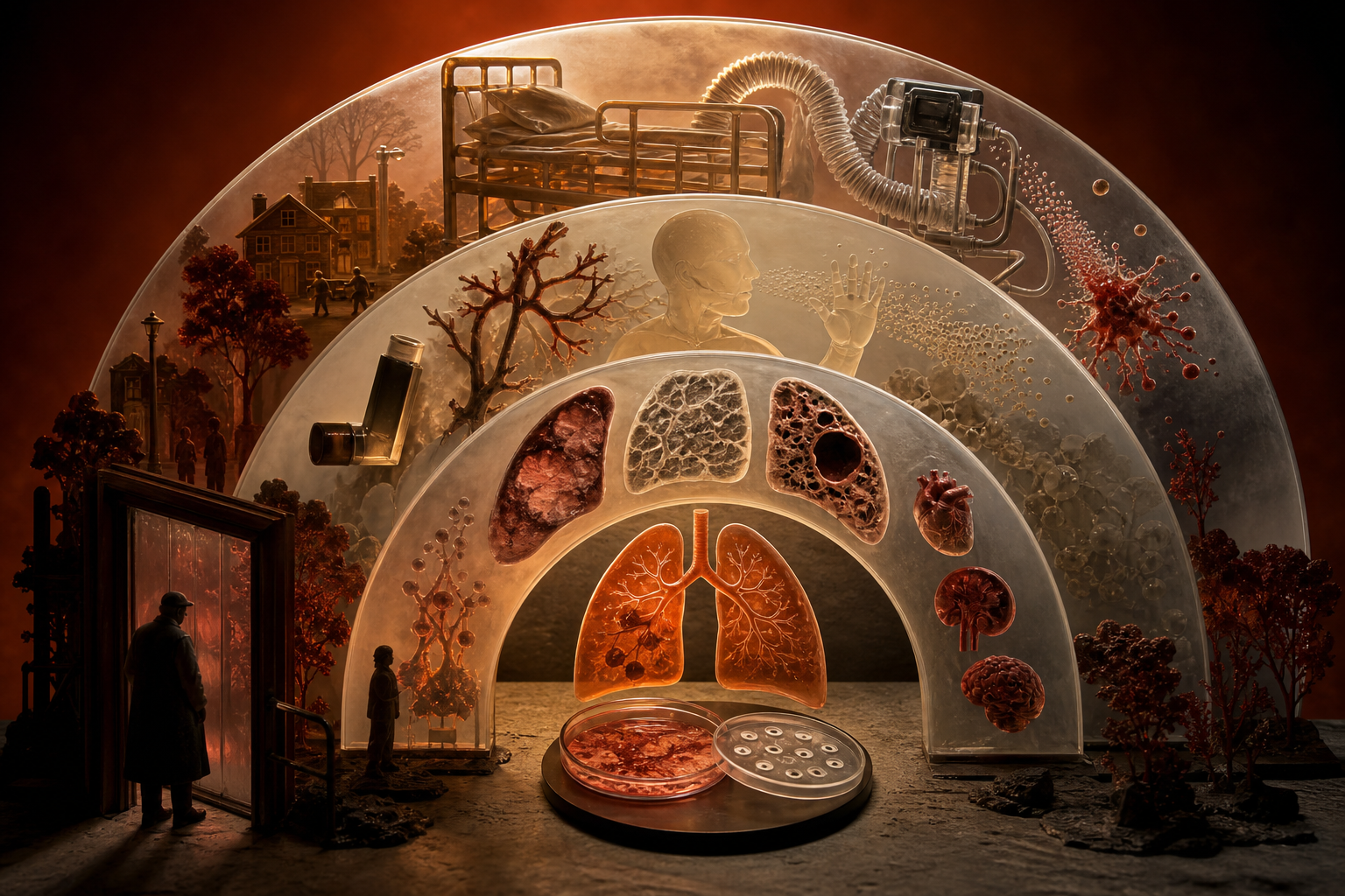
When a vignette asks for the organism, diagnostic test, or initial antibiotic regimen, process the clues in this order:
- **Setting:** Did the illness begin in the community, at least 48 hours after admission, or more than 48 hours after intubation?
- **Host:** Is there COPD, structural lung disease, impaired consciousness, recent influenza, immunosuppression, or prior respiratory colonization?
- **Syndrome:** Is the pattern lobar, interstitial, necrotizing, cavitary, or aspiration dependent? Are gastrointestinal, neurologic, hematologic, or skin findings present?
- **Resistance risk:** Has the patient previously grown MRSA or *Pseudomonas*? Were parenteral antibiotics given during a recent hospitalization? What does the local antibiogram show?
The fourth layer is especially important when the question asks for empiric therapy. The ATS/IDSA community-acquired pneumonia guideline emphasizes prior respiratory isolation and recent hospitalization with parenteral antibiotics rather than using broad healthcare exposure alone to justify MRSA or antipseudomonal treatment. (pmc.ncbi.nlm.nih.gov)
| Clinical setting or host clue | Organisms to prioritize | Findings that support the match | Important trap or treatment implication | |---|---|---|---| | Typical CAP with abrupt fever and focal consolidation | *Streptococcus pneumoniae* | Lobar opacity, pleuritic pain, productive cough; encapsulated gram-positive diplococci | Lobar consolidation is not organism specific | | COPD or older adult with chronic airway disease | Nontypeable *Haemophilus influenzae*, pneumococcus, *Moraxella* | Increased cough, purulent sputum, focal pneumonia or COPD exacerbation | Do not assume every COPD exacerbation is pneumonia | | Young patient in a household, school, barracks, or dormitory | *Mycoplasma pneumoniae* | Gradual fever, persistent dry cough, interstitial or patchy infiltrates, possible hemolysis or rash | Cold agglutinins are suggestive, not confirmatory; β-lactams do not work because the organism lacks a cell wall | | Water-system exposure, recent travel, or severe CAP with gastrointestinal and neurologic findings | *Legionella pneumophila* | Diarrhea, confusion, hyponatremia, elevated liver enzymes | No radiographic pattern is specific; severe disease or outbreak exposure supports urinary antigen plus respiratory testing | | Pneumonia after influenza | *Staphylococcus aureus*, including MRSA | Rapid deterioration, hemoptysis, leukopenia, multilobar disease, cavitation or necrosis | Add MRSA coverage when the overall syndrome and resistance risk justify it | | Cystic fibrosis, bronchiectasis, prior respiratory isolation, HAP, or VAP | *Pseudomonas aeruginosa* and other resistant gram-negative bacilli | Severe illness, structural lung disease, prior cultures, recent IV antibiotics | Green sputum or a “grape-like” odor is trivia, not bedside confirmation | | Alcohol use disorder, diabetes, immunosuppression, or nosocomial exposure | *Klebsiella pneumoniae* and other gram-negative bacilli | Severe consolidation, possible necrosis; classic descriptions include upper-lobe disease and thick bloody sputum | Alcohol use is not specific for *Klebsiella* and also increases aspiration risk | | Loss of consciousness, dysphagia, seizure, or impaired airway protection | Mixed aspiration-associated flora | Dependent-segment infiltrate; abscess may produce foul sputum and an air–fluid level | Routine extra anaerobic coverage is not recommended unless abscess or empyema is suspected |
Community-Acquired Pathogens and Their Decisive Clues
Pneumococcus is the baseline comparison
*Streptococcus pneumoniae* remains the classic reference organism for bacterial CAP. Its polysaccharide capsule limits phagocytosis, while IgA protease supports mucosal colonization. The familiar combination of abrupt fever, pleuritic pain, productive cough, and lobar consolidation should raise its probability.
The mistake is treating “lobar” as synonymous with pneumococcus. *Klebsiella*, *Legionella*, *Haemophilus*, and *Staphylococcus aureus* can also produce airspace consolidation. Imaging establishes the distribution and complications of pneumonia more reliably than it identifies the pathogen.
Mycoplasma combines respiratory and extrapulmonary clues
Think of *Mycoplasma pneumoniae* when a younger patient has an insidious respiratory illness, persistent cough, relatively modest examination findings, and patchy or interstitial opacities. Because *Mycoplasma* has no peptidoglycan cell wall, β-lactams are ineffective; a macrolide or doxycycline is typically used when targeted atypical coverage is required.
Cold agglutinins are IgM antibodies that can accompany infection and occasionally produce hemolysis, but they are neither sensitive nor specific enough to establish the diagnosis. A clinical review of Mycoplasma infection likewise describes the gradual respiratory presentation and the limited specificity of cold-agglutinin testing. (pmc.ncbi.nlm.nih.gov)
Legionella is a multisystem pneumonia
*Legionella pneumophila* is an intracellular gram-negative bacillus associated with aerosolized contaminated water. Pneumonia accompanied by diarrhea, confusion, hyponatremia, transaminase elevation, or renal dysfunction should increase suspicion, especially after travel or a possible outbreak exposure.
These clues remain probabilistic. An authoritative NCBI review of legionellosis diagnosis notes that gastrointestinal, neurologic, and laboratory abnormalities may support the diagnosis, but radiology has no unique Legionella pattern. (ncbi.nlm.nih.gov) For severe CAP, outbreak exposure, or recent travel, guidelines support Legionella urinary antigen testing together with a lower-respiratory culture or nucleic acid test. The urinary antigen primarily detects *L. pneumophila* serogroup 1, so respiratory testing broadens detection.
Post-influenza necrosis points toward Staphylococcus aureus
A patient who improves from influenza and then rapidly develops fever, respiratory failure, hemoptysis, leukopenia, or cavitary infiltrates has a dangerous secondary bacterial pneumonia pattern. *S. aureus*, including MRSA, becomes a major concern.
Some strains produce Panton-Valentine leukocidin, which damages leukocytes and is associated with tissue necrosis. A review of staphylococcal necrotizing pneumonia describes its association with preceding influenza, leukopenia, airway hemorrhage, severe respiratory failure, and rapid progression. (pubmed.ncbi.nlm.nih.gov) The decisive clue is not simply “influenza history”; it is influenza followed by a severe destructive bacterial syndrome.
Klebsiella clues should not overrule the full case
Board resources classically connect *Klebsiella pneumoniae* with alcohol use disorder, diabetes, thick bloody “currant-jelly” sputum, upper-lobe consolidation, and a bulging fissure. The organism’s prominent capsule contributes to virulence and the mucoid appearance of colonies.
However, the classic radiographic package is less dependable than its reputation suggests. In one series of bacteremic cases, modern clinicoradiographic findings did not consistently include cavitation or a bulging fissure. (pubmed.ncbi.nlm.nih.gov) Use alcohol use, diabetes, severe gram-negative disease, and nosocomial exposure as supporting evidence—not proof. In an obtunded patient with poor dentition and an air–fluid level, aspiration-associated lung abscess may fit better.
Aspiration Pneumonitis, Aspiration Pneumonia, and Lung Abscess
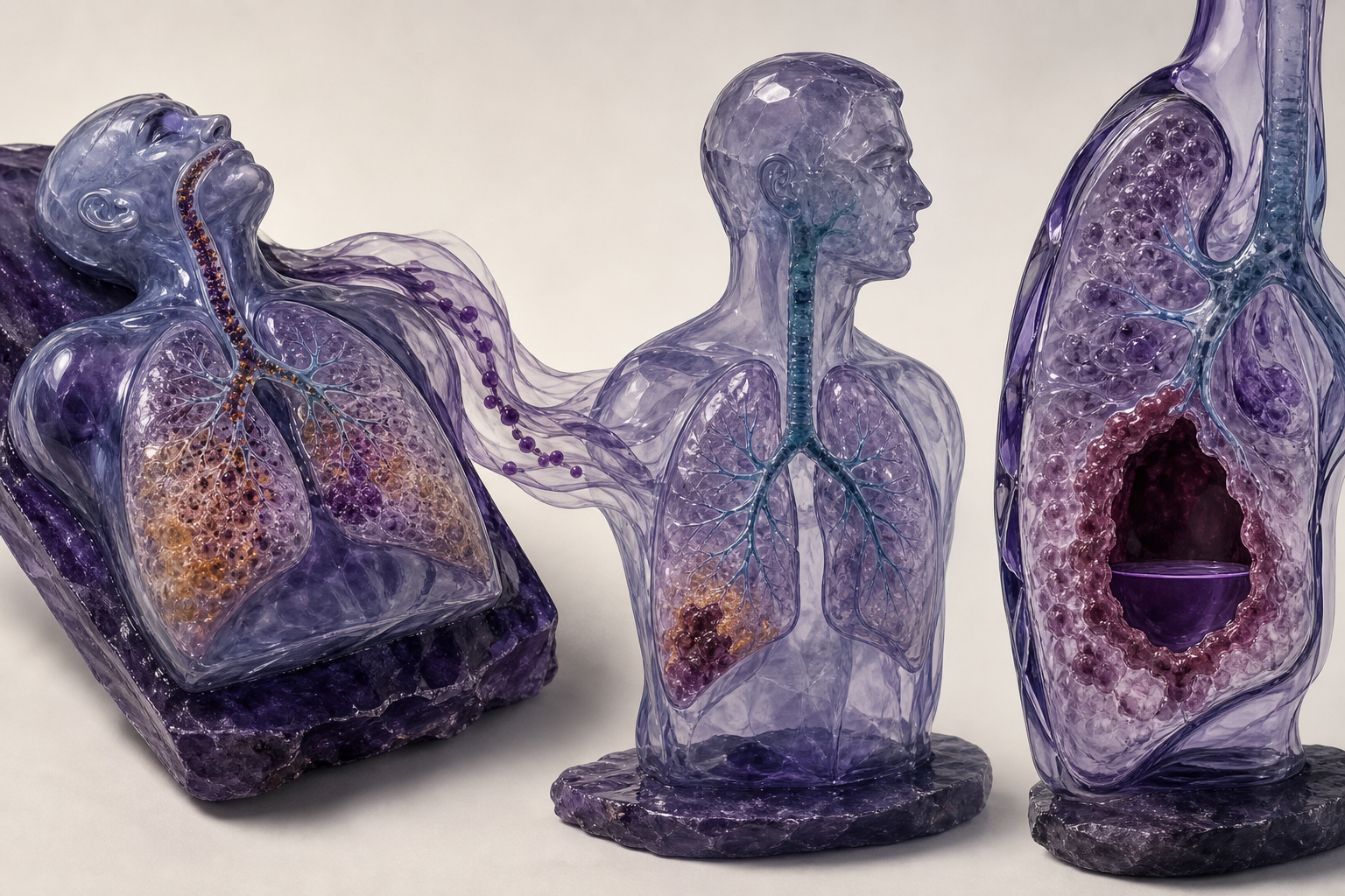
Aspiration questions often test whether antibiotics are needed before they test which antibiotic to use.
**Aspiration pneumonitis** is chemical injury after inhalation of gastric contents. It typically causes abrupt hypoxemia and infiltrates after a witnessed event, and it may improve with supportive care over 24–48 hours. Fever and leukocytosis can occur from inflammation, so they do not automatically prove infection.
**Aspiration pneumonia** is an infection occurring in a patient with impaired airway protection, dysphagia, or recurrent aspiration. The infiltrate often appears in gravity-dependent lung segments: the basal lower lobes when upright and the posterior upper lobes or superior lower lobes when supine.
**Lung abscess** adds parenchymal necrosis and a cavity, often with an air–fluid level, putrid sputum, poor dentition, or a delayed course. This is where anaerobic and mixed oral flora become more important.
The current CAP guideline advises against routinely adding anaerobic therapy for suspected aspiration pneumonia unless lung abscess or empyema is present. (pmc.ncbi.nlm.nih.gov) This corrects the outdated shortcut that every aspiration event requires clindamycin. A contemporary review of aspiration pneumonia also emphasizes standard CAP-directed therapy for most cases and reserves dedicated anaerobic coverage for selected complicated infections. (ncbi.nlm.nih.gov)
How CAP, HAP, and VAP Change Empiric Therapy
Pathogen clues affect probability, but the care setting determines the initial coverage boundary.
For **healthy outpatients with CAP**, guideline-supported choices include amoxicillin or doxycycline; macrolide monotherapy is reserved for areas with sufficiently low pneumococcal macrolide resistance. Outpatients with significant comorbidities generally need a β-lactam plus a macrolide or doxycycline, or a respiratory fluoroquinolone when appropriate. Hospitalized nonsevere CAP is commonly treated with a β-lactam plus a macrolide or with a respiratory fluoroquinolone. Severe CAP requires combination therapy, usually a β-lactam plus a macrolide or respiratory fluoroquinolone. (pmc.ncbi.nlm.nih.gov)
**HAP** begins at least 48 hours after hospital admission and was not incubating at admission. **VAP** begins more than 48 hours after endotracheal intubation. The ATS/IDSA HAP/VAP guideline recommends that empiric therapy reflect local resistance data and cover *S. aureus*, *Pseudomonas*, and other gram-negative bacilli. (pmc.ncbi.nlm.nih.gov)
MRSA coverage with vancomycin or linezolid is added when risk factors or local prevalence warrant it. Two antipseudomonal agents from different classes may be needed when resistance risk or mortality risk is high; otherwise, one active antipseudomonal agent may be sufficient. Obtain respiratory cultures, then narrow therapy when susceptibilities and the clinical course permit.
Worked Vignette: Setting Beats the Flashiest Buzzword
A 67-year-old man is admitted with severe pancreatitis. On hospital day 6, after 72 hours of mechanical ventilation, he develops a temperature of 39.2°C, purulent endotracheal secretions, worsening oxygen requirements, and a new right lower-lobe infiltrate. He received IV ceftriaxone earlier in the admission and now requires norepinephrine. Which empiric regimen is most appropriate while cultures are pending?
**Best answer:** Begin MRSA coverage plus broad antipseudomonal therapy guided by the hospital antibiogram. In a high-mortality patient with prior IV antibiotic exposure, an acceptable conceptual regimen is vancomycin or linezolid plus two antipseudomonal agents from different classes, followed by culture-directed de-escalation.
**Decisive clue:** Pneumonia began more than 48 hours after intubation, making this VAP. Septic shock and prior IV antibiotics increase the consequences of inadequate initial coverage and the probability of resistant organisms.
**Why the nearest distractor loses:** Ceftriaxone plus azithromycin is a standard CAP-style regimen, but it does not reliably cover MRSA or *Pseudomonas*. The right lower-lobe infiltrate might tempt the reader toward aspiration, yet location is less decisive than the ventilator setting, antibiotic exposure, and shock.
For more focused comparisons that connect mechanisms to clinical decisions, explore the CoreStepPrep Core Concepts.
Final Takeaways
- Classify the pneumonia setting before naming an organism or selecting antibiotics.
- Use host factors, extrapulmonary findings, and imaging as probability modifiers—not microbiologic confirmation.
- Think *Legionella* with severe multisystem CAP, *Mycoplasma* with gradual atypical illness, and *S. aureus* with destructive post-influenza disease.
- Do not add routine anaerobic coverage after aspiration unless abscess, empyema, or another strong anaerobic syndrome is present.
- In HAP and VAP, let prior cultures, recent IV antibiotics, illness severity, and the local antibiogram determine MRSA and antipseudomonal coverage.
Try a free USMLE question at CoreStepPrep.