Nephrotic vs Nephritic Syndrome: USMLE Clues
Compare nephrotic and nephritic syndrome by proteinuria, hematuria, urine casts, complement, edema, mechanisms, exceptions, and USMLE clues.
Nephrotic syndrome is primarily a **protein-leak syndrome**, whereas nephritic syndrome is primarily an **inflammatory glomerular bleeding syndrome**. On a Step 1 vignette, prioritize the urine sediment: heavy proteinuria with fatty casts suggests nephrotic disease, while dysmorphic red cells or red blood cell casts strongly support nephritic disease—even when edema and proteinuria appear in both.
Nephrotic vs Nephritic Syndrome at a Glance
The terms *nephrotic* and *nephritic* describe clinical patterns, not single diseases. Nephrotic syndrome classically combines nephrotic-range proteinuria—more than 3.5 g/day in adults—with hypoalbuminemia, edema, and hyperlipidemia. Nephritic syndrome combines glomerular hematuria with variable proteinuria, hypertension, fluid retention, and often reduced kidney function.
| Finding | Nephrotic syndrome | Nephritic syndrome | Important exception | |---|---|---|---| | Proteinuria | Usually heavy; often >3.5 g/day | Usually subnephrotic | IgA nephropathy, proliferative lupus nephritis, and MPGN can produce nephrotic-range proteinuria | | Hematuria | Usually absent or microscopic | Prominent; may be cola-colored | FSGS and membranous nephropathy may cause microscopic hematuria | | Urine casts | Fatty casts and oval fat bodies | RBC casts; sometimes mixed cellular casts | RBC casts may be absent despite active glomerulonephritis | | Serum complement | Usually normal | May be low or normal depending on cause | Complement identifies the mechanism; it does not define the syndrome | | Edema | Often marked or generalized | Usually mild to moderate | Severe nephritic disease can cause substantial volume overload | | Blood pressure | Normal or elevated | Frequently elevated | Sodium retention may raise blood pressure in either syndrome | | Kidney function | May initially be preserved | Creatinine often rises | Severe nephrotic disease can also cause acute kidney injury | | Serum lipids | Frequently elevated | Not a defining feature | Mixed syndromes may show both inflammatory and nephrotic findings |
These are tendencies rather than absolute borders. Modern clinicopathologic reviews emphasize that nephrotic and nephritic presentations occupy opposite ends of a spectrum, with lupus nephritis and membranoproliferative glomerulonephritis among the important causes of a mixed pattern (Patterns of Glomerular Injury).
Why the Urine Looks Different: Barrier Leak vs Inflammation
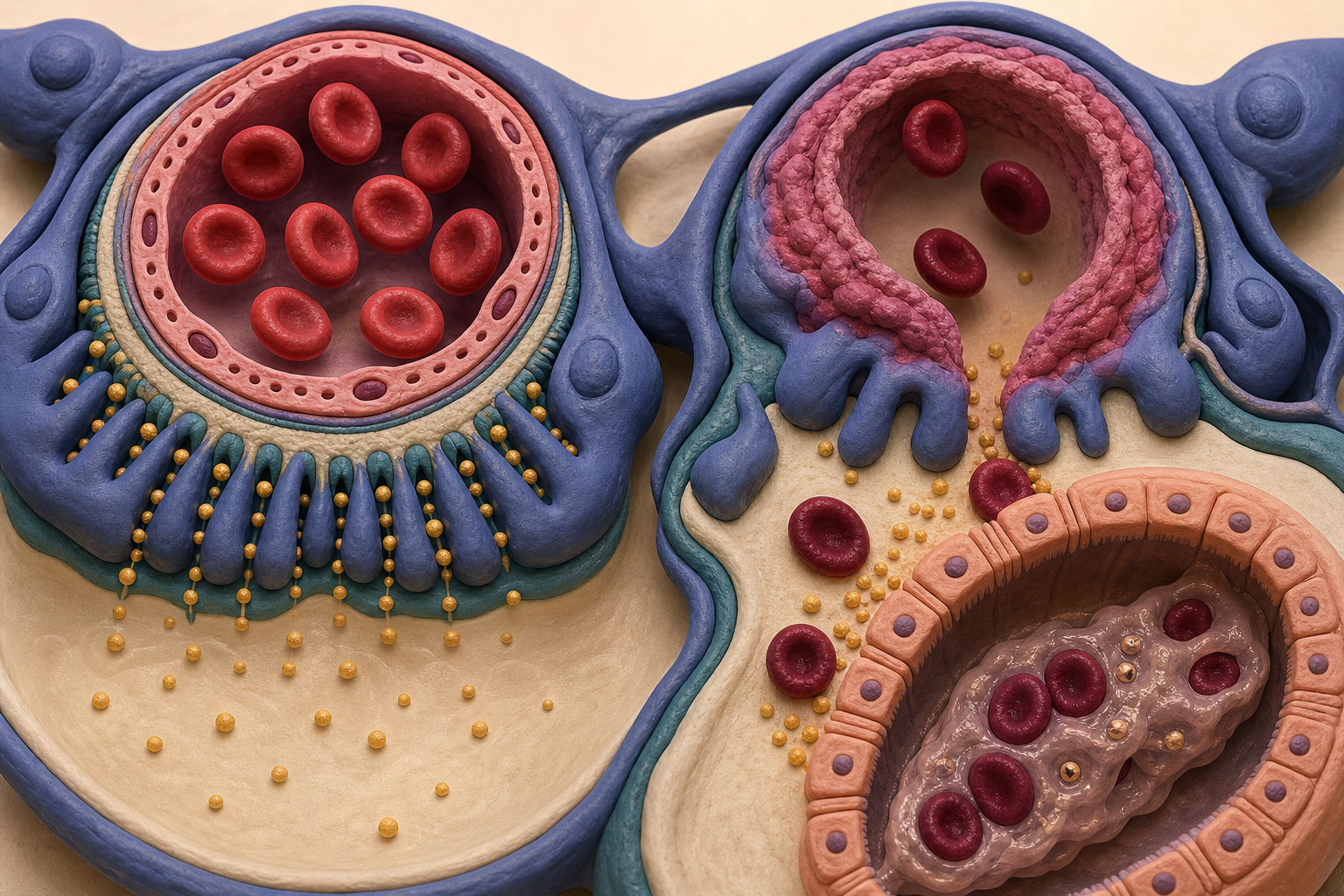
The glomerular filtration barrier contains fenestrated endothelial cells, the glomerular basement membrane, and podocyte foot processes joined by slit diaphragms. Together, these structures restrict plasma proteins according to molecular size, charge, and shape (Properties of the Glomerular Barrier and Mechanisms of Proteinuria).
In **nephrotic syndrome**, podocyte or capillary-wall injury increases protein permeability. Albumin enters the urine, serum albumin falls, and edema develops through a combination of reduced plasma oncotic pressure and renal sodium retention. The liver increases lipoprotein production, contributing to hyperlipidemia; filtered lipids can produce oval fat bodies and fatty casts.
For Step 1, the traditional model describes loss of charge selectivity as an explanation for albuminuria, especially in minimal change disease. That model remains useful, but it should not be interpreted as the basement membrane’s negative charge acting alone. Current physiology treats the filtration barrier as a dynamic, integrated structure in which the endothelium, basement membrane, podocytes, and tubular albumin handling all contribute.
In **nephritic syndrome**, inflammatory injury disrupts glomerular capillary walls and permits red cells to enter Bowman space. The cells become distorted while crossing the damaged barrier, producing dysmorphic RBCs. When red cells become trapped in a tubular protein matrix, they form RBC casts—evidence that the bleeding originated within the nephron rather than from the bladder or ureter.
Inflammatory swelling and cellular proliferation also reduce filtration. The resulting fall in glomerular filtration rate promotes sodium and water retention, explaining the combination of oliguria, hypertension, azotemia, and edema.
Proteinuria, Hematuria, and Casts: The Highest-Yield Sequence
When a vignette provides many renal findings, interpret them in this order.
1. Decide whether the urine sediment is active
An **active sediment** contains findings such as dysmorphic RBCs, RBC casts, or mixed cellular casts. This points toward inflammatory glomerular injury and therefore a nephritic process.
A **bland sediment** lacks substantial cellular activity. In a patient with marked proteinuria, hypoalbuminemia, and edema, a bland sediment favors a podocyte-predominant nephrotic process.
2. Quantify the protein loss
Heavy proteinuria strongly favors nephrotic syndrome, especially when paired with low serum albumin and hyperlipidemia. However, protein quantity alone cannot exclude nephritic disease. IgA nephropathy and proliferative lupus nephritis may produce both active sediment and nephrotic-range protein loss.
Conversely, blood on urinalysis does not automatically make a process nephritic. Microscopic hematuria can accompany podocytopathies, including minimal change disease, FSGS, and membranous nephropathy (Hematuria in Podocytopathies). The stronger nephritic clue is **glomerular hematuria**, particularly dysmorphic RBCs or RBC casts.
3. Identify the cast type
Urine casts localize pathology because they form in renal tubules:
- **RBC casts:** glomerulonephritis and a nephritic sediment
- **Fatty casts or oval fat bodies:** nephrotic-range proteinuria
- **WBC casts:** pyelonephritis, acute interstitial nephritis, or inflammatory glomerular disease
- **Muddy brown granular casts:** acute tubular injury
Fat droplets containing cholesterol may display a Maltese-cross pattern under polarized light. RBC casts are highly suggestive of nephritic disease, but their absence does not rule it out because casts may be sparse or missed during specimen examination. A review of urine microscopy identifies RBC casts as a distinguishing nephritic finding and fatty casts as a marker of nephrotic-range proteinuria (Diagnostic Utility of Urine Microscopy).
Complement Narrows the Nephritic Differential
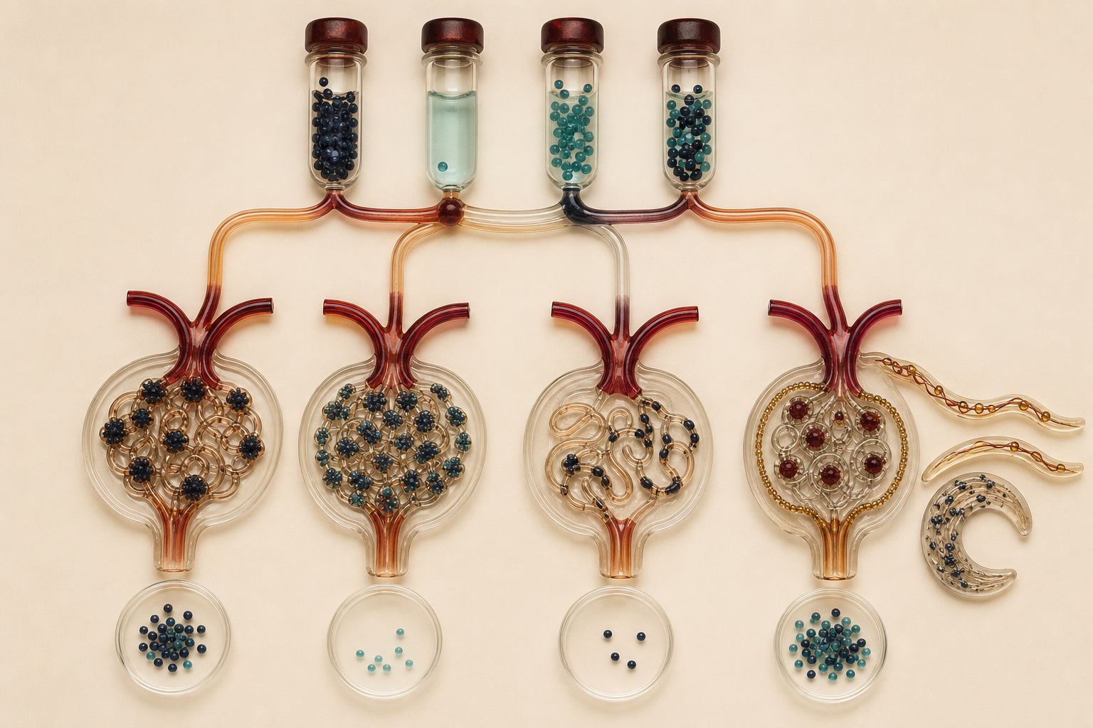
Complement is not simply “low in nephritic syndrome.” It is normal in several major nephritic diseases, so the better question is: **Which inflammatory mechanism is consuming complement?**
| Complement pattern | Important associations | Supporting clue | |---|---|---| | Low C3, often normal C4 | Postinfectious GN, C3 glomerulopathy | Recent infection or C3-dominant deposits | | Low C3 and low C4 | Lupus nephritis; sometimes endocarditis-associated GN | Systemic autoimmune or infectious findings | | Low C4 with relatively preserved C3 | Cryoglobulinemic GN | Purpura, neuropathy, arthralgia, hepatitis association | | Usually normal C3 and C4 | IgA nephropathy, ANCA-associated GN, anti-GBM disease | Disease-specific timing, antibodies, or pulmonary findings |
Serum complement levels and renal complement deposition are related but not interchangeable. Complement can be activated within the kidney without producing low circulating C3 or C4. Still, the serum pattern is a valuable way to narrow the nephritic differential; a review of complement-mediated kidney disease summarizes the characteristic patterns for postinfectious GN, lupus nephritis, IgA nephropathy, ANCA-associated disease, and anti-GBM disease (The Role of Complement in Kidney Disease).
Edema Occurs in Both Syndromes for Different Reasons
Marked edema does not automatically mean nephrotic syndrome.
In nephrotic syndrome, urinary albumin loss lowers plasma oncotic pressure, favoring movement of water into the interstitial space. Reduced effective circulating volume may activate sodium-retaining pathways, while some patients also demonstrate primary renal sodium retention. The result can be dependent edema, periorbital swelling, ascites, or anasarca.
In nephritic syndrome, inflammation reduces the filtration surface and GFR. Sodium and water accumulate intravascularly, making hypertension, pulmonary congestion, and oliguria particularly useful companion clues. Periorbital edema can still be prominent, especially in children.
Thus, pair edema with hemodynamics:
- **Generalized edema + profound hypoalbuminemia + heavy proteinuria:** nephrotic pattern
- **Edema + hypertension + oliguria + rising creatinine:** nephritic pattern
The systemic consequences also differ. Nephrotic syndrome can increase the risks of thrombosis and infection because anticoagulant and immune proteins may be lost in urine; hyperlipidemia is another characteristic laboratory consequence (MedlinePlus Nephrotic Syndrome).
Disease Patterns Worth Recognizing
Classic nephrotic diseases include:
- **Minimal change disease:** diffuse podocyte foot-process effacement; often steroid responsive
- **FSGS:** focal and segmental sclerosis; associated with causes such as HIV, obesity, heroin exposure, and sickle cell disease
- **Membranous nephropathy:** subepithelial immune deposits with capillary-wall thickening
- **Diabetic kidney disease:** mesangial expansion and nodular glomerulosclerosis
- **Amyloidosis:** Congo red-positive deposits with apple-green birefringence
Classic nephritic diseases include:
- **Postinfectious glomerulonephritis:** immune-complex disease, commonly with low C3
- **IgA nephropathy:** hematuria occurring close to a respiratory or gastrointestinal infection; complement usually normal
- **Proliferative lupus nephritis:** active sediment with low C3 and C4
- **ANCA-associated glomerulonephritis:** pauci-immune crescentic disease, usually with normal complement
- **Anti-GBM disease:** rapidly progressive GN, sometimes with pulmonary hemorrhage
Subepithelial injury tends to produce protein leakage with less glomerular inflammation, whereas subendothelial deposits are exposed to circulating inflammatory mediators and more readily generate a nephritic pattern. This anatomic principle explains why membranous nephropathy is usually nephrotic while proliferative lupus nephritis is commonly nephritic or mixed.
Worked Vignette: The Decisive Clue Is the Sediment
A 17-year-old boy develops facial swelling and dark urine two weeks after a crusted skin infection. His blood pressure is 154/96 mm Hg. Laboratory testing shows a serum creatinine of 2.0 mg/dL, low C3, normal C4, and 1.8 g of urinary protein per day. Urine microscopy shows dysmorphic RBCs and RBC casts.
**Most likely pattern:** nephritic syndrome due to postinfectious glomerulonephritis.
The decisive clue is the **RBC cast**, which localizes bleeding to an inflamed glomerulus. Hypertension, rising creatinine, subnephrotic proteinuria, and low C3 reinforce that interpretation.
**Why the nearest distractor loses:** Minimal change disease could explain facial edema after an immune trigger, but it should produce dominant heavy proteinuria, hypoalbuminemia, and a relatively bland or lipid-rich sediment. It does not explain RBC casts, reduced GFR, hypertension, and complement consumption as a unified pattern.
Notice that edema was not the deciding feature. The sediment, kidney function, protein quantity, and complement pattern formed the diagnostic chain.
Management Follows the Syndrome but Targets the Cause
Initial management addresses dangerous consequences—volume overload, severe hypertension, electrolyte abnormalities, acute kidney injury, thrombosis, or infection—while the underlying disease is identified.
For nephrotic disease, supportive care may include sodium restriction, diuretics for edema, blood-pressure and proteinuria reduction, and treatment of dyslipidemia or thrombosis risk when clinically indicated. Disease-specific therapy depends on whether the lesion is minimal change disease, FSGS, membranous nephropathy, diabetes, amyloidosis, or another cause.
For nephritic disease, the urgency depends on the rate of kidney-function loss and systemic findings. Rapidly rising creatinine, RBC casts, pulmonary hemorrhage, or suspected crescentic glomerulonephritis requires prompt evaluation. Therapy differs substantially among infection-related GN, lupus nephritis, ANCA-associated vasculitis, and anti-GBM disease, so “treat nephritic syndrome” is never a sufficient final diagnosis.
For related renal mechanisms and pathology comparisons, explore the CoreStepPrep Core Concepts.
Final Takeaways
- Nephrotic syndrome is dominated by heavy protein loss; nephritic syndrome is dominated by inflammatory glomerular hematuria.
- RBC casts and dysmorphic RBCs outweigh edema as evidence of a nephritic process.
- Fatty casts, hypoalbuminemia, and hyperlipidemia support nephrotic syndrome.
- Complement narrows the cause of nephritic disease but may be either low or normal.
- Mixed presentations are real: interpret the entire pattern rather than relying on one absolute rule.
Try a free USMLE question at CoreStepPrep