Types I–IV Hypersensitivity: Mechanisms and Examples
Compare types I–IV hypersensitivity by immune mediator, timing, and disease examples. Distinguish fixed targets from immune complexes with a worked vignette.
Types I–IV hypersensitivity reactions differ primarily by **what causes the injury**: type I uses IgE-triggered mast cells; type II uses antibodies against fixed cell or tissue targets; type III uses deposited antigen–antibody complexes; and type IV uses T cells. Timing helps, but the decisive distinction is the immune mechanism—not simply whether symptoms appear immediately or several days later. The classical hypersensitivity classification separates these mechanisms even when their clinical findings overlap.
Types I–IV hypersensitivity: mediators, timing, and examples
Start with three questions: Is antibody involved? If so, which class? And does the antibody recognize a fixed target, or form complexes with soluble antigen? These questions distinguish the four mechanisms more reliably than a symptom such as rash or swelling. The FDA’s immunotoxicity guidance identifies IgE-mediated type I, IgG/IgM-associated types II and III, and T-cell–mediated type IV reactions.
| Type | Defining mechanism | Principal effectors | Characteristic timing | Contrasting examples | |---|---|---|---|---| | **I: immediate** | Allergen cross-links mast-cell–bound IgE | Mast cells, histamine, lipid mediators; eosinophils in the late phase | Usually minutes after re-exposure; later inflammation can follow | IgE-mediated anaphylaxis, allergic rhinitis, allergic asthma | | **II: antibody-mediated** | IgG or IgM recognizes a fixed cell-surface or extracellular-matrix antigen | Phagocytes, complement, or altered receptor signaling | Variable; minutes to hours with preexisting antibody, or ongoing autoimmune disease | Immune hemolytic anemia, anti-GBM disease, Graves disease | | **III: immune complex–mediated** | Antibody binds soluble antigen; complexes deposit or form in tissues | Complement and recruited inflammatory cells, especially neutrophils | Hours for an Arthus reaction; roughly 1–2 weeks for initial serum sickness | Arthus reaction, serum sickness, poststreptococcal glomerulonephritis | | **IV: T-cell–mediated** | Sensitized T cells cause inflammation or kill target cells | CD4⁺ T cells, macrophages, CD8⁺ cytotoxic T cells | Classically develops over 24–72 hours after re-exposure | Allergic contact dermatitis, tuberculin skin reaction |
The table summarizes antibody-dependent mechanisms, T-cell–mediated hypersensitivity, and the disease-specific timing discussed below. These intervals are patterns, not rigid diagnostic cutoffs.
Type I: IgE equips mast cells for rapid activation
Type I begins with **sensitization**. Allergen-specific helper T cells support B-cell class switching to IgE through IL-4/IL-13 signaling and CD40–CD40L interaction. The resulting IgE binds the high-affinity receptor **FcεRI** on mast cells, preparing them to respond to subsequent exposure. This sequence is described in the NCBI immunobiology reference on IgE production.
On re-exposure, an allergen cross-links adjacent IgE molecules. Mast cells release preformed mediators, including histamine, and generate lipid mediators such as leukotrienes and prostaglandins. Increased vascular permeability produces swelling and urticaria; airway smooth-muscle contraction contributes to bronchospasm. A later inflammatory phase recruits eosinophils and other leukocytes. Importantly, **late-phase type I inflammation is not type IV hypersensitivity**: the initiating mechanism remains IgE-mediated mast-cell activation. See the mechanisms of allergic effector responses.
The clearest clinical contrast is localized allergy versus systemic anaphylaxis. Sneezing after pollen exposure and hypotension with wheezing after a food exposure can share an initiating mechanism, but differ greatly in distribution and severity. For anaphylaxis, prompt intramuscular epinephrine is first-line treatment; antihistamines are adjuncts, not substitutes for treating respiratory or circulatory compromise, according to CDC anaphylaxis management guidance.
Type II: antibodies recognize a fixed target
The defining feature of type II is **where the antigen is located**: on a cell surface or within extracellular matrix. Antibody binding can recruit complement and inflammatory cells, promote clearance of the targeted cell, or alter receptor function. Thus, “antibody-mediated” is more accurate than treating every type II reaction as purely cytotoxic. The classical immunobiology classification distinguishes these fixed targets from the antigen–antibody complexes of type III.
Cell injury versus receptor dysfunction
In immune hemolytic anemia, antibodies target red cells and promote their premature destruction. This is a cell-directed process, not injury from circulating complexes lodging in unrelated tissues. The MedlinePlus description of immune hemolytic anemia connects antibody targeting with red-cell destruction and laboratory evidence of hemolysis.
A direct antiglobulin, or direct Coombs, test detects antibody and/or complement coating the patient’s red cells, depending on the reagent used. That makes the test a useful mechanistic clue: the immune reactants are attached to the affected cell. The principle is documented in FDA-hosted antiglobulin reagent instructions.
Receptor-directed disease illustrates why cell destruction is not required:
- **Graves disease:** stimulating antibodies activate the TSH receptor and increase thyroid hormone production, as explained by NIDDK’s Graves disease resource.
- **Myasthenia gravis:** antibodies interfere with neuromuscular transmission; most affected patients have antibodies against the acetylcholine receptor, while some have antibodies against related proteins such as MuSK. MedlinePlus Genetics describes the resulting reduction in available acetylcholine receptors.
These are contrasting outcomes of antibody binding: excessive receptor stimulation versus impaired signaling.
Type II versus type III: fixed targets or deposited complexes?
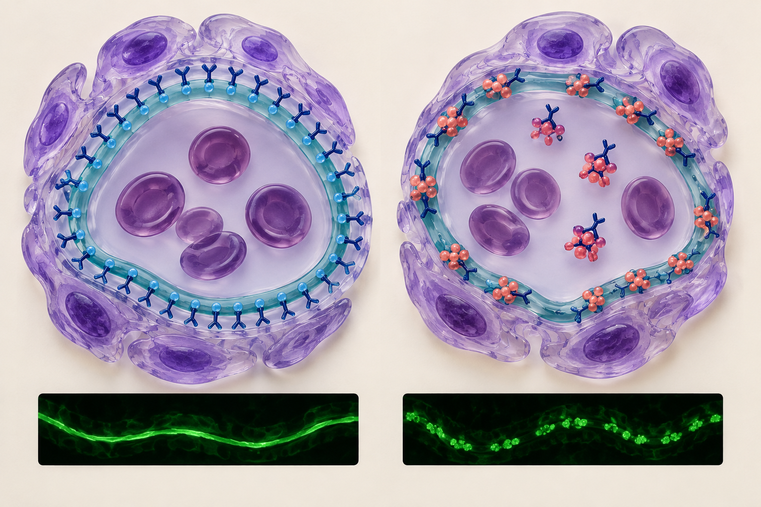
Both types can involve IgG and complement. The distinction is not “antibody versus complement”; it is **direct recognition of tissue versus immune-complex deposition**. A useful comparison is anti-GBM disease against immune-complex glomerulonephritis, as described in the renal pathology consensus classification.
In anti-GBM disease, antibodies directly attack basement-membrane targets in the kidneys and sometimes the lungs. This can produce glomerulonephritis and pulmonary hemorrhage. The fixed tissue target makes it type II; NIDDK’s anti-GBM disease explanation describes this antibody attack.
On renal immunofluorescence, anti-GBM disease characteristically produces **linear IgG staining** along the glomerular basement membrane. Immune-complex glomerulonephritis typically produces **granular deposits** instead. Think of antibody tracing a continuous structural target versus complexes appearing in discontinuous deposits. These patterns are specified in the renal pathology consensus report.
Type III: immune complexes recruit complement and neutrophils
In type III, antibodies bind soluble antigens. Complexes that deposit or form in tissues activate complement and recruit inflammatory cells, producing tissue injury. A localized example is an **Arthus reaction**: immune complexes form near an antigen injection site in someone with substantial preexisting antibody. It typically causes painful local swelling several hours later, rather than immediate systemic anaphylaxis. CDC’s description of Arthus reactions identifies local vasculitis, immune-complex deposition, complement activation, and a characteristic 4–12-hour onset.
**Serum sickness** is the systemic contrast. After exposure to a foreign protein, patients may develop fever, rash, arthralgia, and renal involvement as immune complexes form. In a prospective clinical and immunologic study of serum sickness, symptoms appeared 8–13 days after horse antithymocyte globulin treatment, circulating complexes developed, and C3/C4 levels fell.
Do not assume that every immune-complex disease lowers C3 and C4 identically. Poststreptococcal glomerulonephritis commonly features low C3 and follows streptococcal infection after a latent interval—approximately 10 days after pharyngitis or up to 3 weeks after a skin infection. Hematuria, edema, hypertension, and red-cell casts support the nephritic presentation described in CDC’s PSGN guidance.
Type IV: T cells produce delayed inflammation or cytotoxicity
Type IV does not require antibody as its defining effector. CD4⁺ T cells can release cytokines that recruit and activate macrophages; CD8⁺ T cells can directly injure target cells. In classic delayed hypersensitivity, IFN-γ links T-cell recognition to macrophage activation. Allergic contact dermatitis and tuberculin reactions illustrate these T-cell–mediated mechanisms.
A tuberculin skin reaction is a useful timing anchor. Previously sensitized T cells recognize the injected antigen and generate local inflammation. The reaction is read at **48–72 hours**, and the measured finding is **induration, not redness**, according to CDC tuberculin skin-test guidance.
Allergic contact dermatitis, such as a delayed eruption after nickel exposure, should also be distinguished from **irritant** contact dermatitis. Irritant dermatitis reflects direct skin injury rather than an antigen-specific delayed immune response. Avoiding the trigger and using topical corticosteroids when appropriate are common management measures described by MedlinePlus’s contact dermatitis resource.
Worked vignette: a delayed rash does not automatically mean type IV
**Original teaching vignette:** A patient receives horse-derived antithymocyte globulin for aplastic anemia. Ten days later, fever, a widespread rash, and painful joints develop. Testing shows reduced C3 and C4 and new proteinuria. Which mechanism best explains the findings?
A. Allergen cross-linking of mast-cell–bound IgE B. Antibodies binding a fixed cell-surface antigen C. Antigen–antibody complexes activating complement in tissues D. Sensitized T cells directly injuring epidermal cells
**Answer: C—type III hypersensitivity.**
The decisive clue is the combination of **foreign-protein exposure, delayed multisystem illness, and complement consumption**. The rash alone does not classify the reaction. This pattern is consistent with the immune complexes and complement changes documented in the prospective serum sickness study.
The nearest distractor is D because the reaction is delayed. However, T-cell–mediated epidermal injury does not best unify the arthralgia, proteinuria, and complement consumption. A is the immediate IgE mechanism; B requires an identified fixed cellular or tissue target. This is a worked application of the mechanism-based classification, not a timing-only diagnosis.
Final takeaways
These distinctions follow the classical hypersensitivity framework:
- **Type I:** IgE activates mast cells; a later inflammatory phase does not change its classification.
- **Type II:** antibodies recognize fixed targets and can destroy cells or change receptor function.
- **Type III:** antigen–antibody complexes cause inflammatory injury; systemic complement consumption can support the diagnosis.
- **Type IV:** T cells drive inflammation or cytotoxicity. Delayed onset alone is insufficient to identify it.
For related explanations, explore the CoreStepPrep core concepts library. Then apply the distinction to a clinical stem: Try a free USMLE question at CoreStepPrep.
Sources and further reading
- www.ncbi.nlm.nih.gov/books/NBK10759/?lang=en-gb
- www.fda.gov/regulatory-information/search-fda-guidance-documents/immunotoxicity-testing-guidance
- www.ncbi.nlm.nih.gov/books/NBK10756
- www.ncbi.nlm.nih.gov/sites/books/NBK27136
- www.ncbi.nlm.nih.gov/books/NBK27117
- www.ncbi.nlm.nih.gov/sites/books/NBK27112
- www.cdc.gov/vaccines/hcp/imz-best-practices/preventing-managing-adverse-reactions.html
- medlineplus.gov/ency/article/000576.htm
- www.fda.gov/media/110737/download
- www.niddk.nih.gov/health-information/endocrine-diseases/graves-disease
- www.medlineplus.gov/genetics/condition/myasthenia-gravis
- pmc.ncbi.nlm.nih.gov/articles/PMC4849835
- www.niddk.nih.gov/health-information/kidney-disease/glomerular-disease/anti-gbm-goodpastures-disease
- www.cdc.gov/MMWR/Preview/MMWRhtml/rr5517a1.htm
- pubmed.ncbi.nlm.nih.gov/6387492
- www.cdc.gov/group-a-strep/hcp/clinical-guidance/post-streptococcal-glomerulonephritis.html
- www.cdc.gov/tb/hcp/testing-diagnosis/tuberculin-skin-test.html
- medlineplus.gov/ency/article/000869.htm