HCM vs Aortic Stenosis: Murmur Maneuvers
Learn why Valsalva and standing amplify HCM but soften aortic stenosis, while squatting reverses the pattern, with a USMLE vignette and table.
Hypertrophic cardiomyopathy (HCM) gets louder when preload falls because a smaller left ventricular cavity worsens its **dynamic** outflow obstruction. Aortic stenosis (AS) usually gets softer when preload falls because less blood crosses its **fixed valvular** obstruction; squatting reverses this pattern by increasing ventricular filling and forward flow.
That mechanism—not a memorized list—is the key to distinguishing HCM from aortic stenosis during USMLE murmur maneuvers.
The core distinction: dynamic obstruction versus fixed valve narrowing
Both HCM and aortic stenosis can produce a harsh crescendo–decrescendo systolic murmur. The crucial difference is what creates the obstruction.
In obstructive HCM, hypertrophy of the interventricular septum narrows the left ventricular outflow tract. During systole, anterior movement of the mitral valve toward the septum—systolic anterior motion, or SAM—further narrows the outflow tract. The degree of obstruction can therefore change from beat to beat as ventricular volume and contractility change.
HCM is commonly associated with pathogenic variants in sarcomeric protein genes, especially *MYH7* and *MYBPC3*, and familial disease is typically inherited in an autosomal dominant pattern. The resulting phenotype can include asymmetric septal hypertrophy, myocyte disarray, interstitial fibrosis, impaired diastolic relaxation, and ventricular arrhythmias. The genetics and variable clinical expression are summarized by MedlinePlus Genetics.
In aortic stenosis, the narrowed structure is the aortic valve itself. The valve opening does not become anatomically tighter simply because the ventricle contains less blood. However, the murmur remains **flow dependent**: less forward flow across the stenotic valve produces less turbulence and a softer murmur. A fixed lesion does not mean fixed murmur intensity.
Why preload separates HCM from aortic stenosis
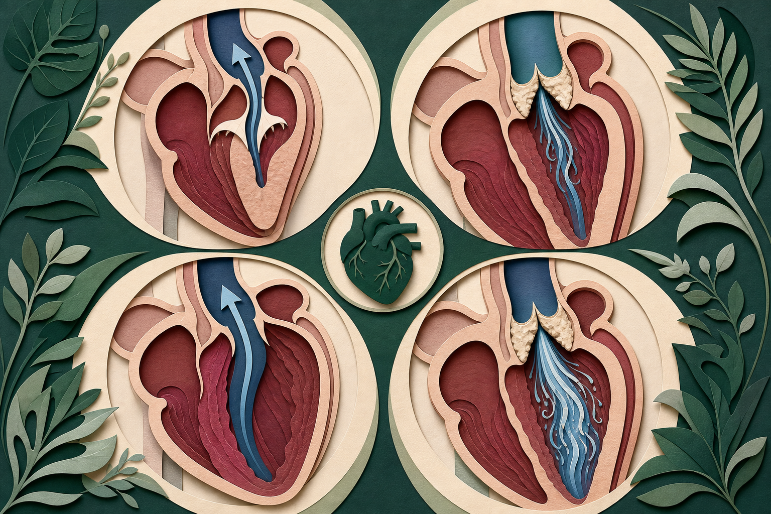
Preload determines how much blood fills the ventricle before contraction. Standing suddenly or performing the strain phase of Valsalva reduces venous return, so left ventricular end-diastolic volume falls.
HCM: a smaller chamber worsens the obstruction
With less blood in the left ventricle, the cavity becomes smaller. The hypertrophied septum and anterior mitral leaflet are brought closer together, SAM becomes more pronounced, and the dynamic LV outflow tract becomes narrower. Blood accelerates through that narrowed tract, so the HCM murmur becomes louder.
This creates the classic sequence:
**Lower preload → smaller LV cavity → more LVOT obstruction → louder HCM murmur**
Standing and Valsalva strain therefore accentuate obstructive HCM. Squatting increases venous return and systemic vascular resistance, enlarging the ventricular cavity and opposing rapid ejection; the obstruction and murmur diminish. These responses are described in a clinical review of HCM auscultation and have also been demonstrated with simultaneous auscultation and Doppler measurements during squatting in patients with obstructive HCM in JACC Case Reports.
Aortic stenosis: lower flow softens the murmur
During Valsalva strain or standing, reduced preload means less blood is ejected across the stenotic aortic valve. The valve remains narrowed, but the lower transvalvular flow produces less turbulence. The murmur becomes softer.
Squatting has the opposite effect. Increased venous return raises ventricular filling and stroke volume, sending more blood through the stenotic valve and usually making the murmur louder.
The corresponding sequence is:
**Lower preload → less transvalvular flow → softer aortic stenosis murmur**
A review of valvular aortic stenosis specifically contrasts its softer murmur during Valsalva with the louder murmur of dynamic HCM.
HCM vs aortic stenosis maneuver table
| Maneuver | Main hemodynamic change | HCM murmur | Aortic stenosis murmur | Mechanistic explanation | |---|---|---:|---:|---| | **Standing from supine** | ↓ Preload | **Louder** | **Softer** | A smaller LV worsens dynamic HCM obstruction, while reduced flow softens AS | | **Squat-to-stand** | Rapid ↓ preload | **Louder** | **Softer** | Same principle as standing, often with a more noticeable transition | | **Valsalva strain** | ↓ Venous return and preload | **Louder** | **Softer** | HCM obstruction increases as the LV empties; AS receives less forward flow | | **Squatting** | ↑ Preload and afterload | **Softer** | **Louder** | A fuller LV relieves HCM obstruction; increased stroke volume raises flow across AS | | **Passive leg raise** | ↑ Venous return | **Softer** | Usually **louder** | Increased filling enlarges the HCM cavity and increases flow across the aortic valve | | **Valsalva release** | Venous return rebounds | **Softer** | **Louder after several beats** | Restored filling reduces HCM obstruction and restores flow through AS | | **Sustained handgrip** | ↑ Afterload | Usually **softer** | Often **softer or minimally changed** | Higher afterload reduces rapid ejection in HCM but may also reduce forward flow across AS |
**Best discriminator:** use Valsalva strain, standing, or squat-to-stand. Handgrip is less useful for separating these two lesions because it may reduce both murmurs. Handgrip is more valuable when comparing an ejection murmur with regurgitant lesions such as mitral regurgitation, which typically becomes louder as afterload rises.
Auscultation and pulse findings that reinforce the diagnosis
Do not make the diagnosis from a single maneuver if the vignette provides additional physical findings.
| Feature | Hypertrophic cardiomyopathy | Aortic stenosis | |---|---|---| | **Typical location** | Left lower sternal border or between the left sternal border and apex | Right upper sternal border | | **Radiation** | Usually not prominent in the carotids | Classically radiates to the carotids | | **Carotid pulse** | Brisk; may have a bifid “spike-and-dome” contour | Delayed and diminished, or *pulsus parvus et tardus* | | **S4** | Common because the hypertrophied LV is stiff | May occur with significant LV hypertrophy | | **Valsalva strain** | Louder | Softer | | **Squatting** | Softer | Louder | | **Typical structural lesion** | Asymmetric septal hypertrophy with possible SAM | Narrowed, abnormal aortic valve |
An S4 reflects atrial contraction into a noncompliant ventricle, so it is not exclusive to HCM. Likewise, exertional syncope, dyspnea, and angina can occur in either condition. The maneuver response, murmur location, radiation, and carotid pulse must be integrated.
One useful mental model is to ask where the bottleneck sits:
- **HCM:** the bottleneck is a muscular tunnel whose width changes with chamber size.
- **Aortic stenosis:** the bottleneck is the valve, while murmur intensity changes primarily with the amount of blood crossing it.
For additional mechanism-first reviews, explore the CoreStepPrep Core Concepts.
Worked vignette: identifying the decisive maneuver
A 19-year-old man develops lightheadedness near the end of basketball practice. His maternal uncle died suddenly at age 31. Cardiac examination reveals a grade 3/6 crescendo–decrescendo systolic murmur heard best along the left lower sternal border. The murmur becomes louder when the patient stands and softer when he squats. A fourth heart sound is present.
Which mechanism most directly explains the change in murmur intensity when he stands?
A. Increased venous return raises flow across a stenotic aortic valve B. Reduced left ventricular volume worsens dynamic outflow tract obstruction C. Increased systemic vascular resistance raises mitral regurgitant volume D. Reduced right ventricular filling delays pulmonic valve closure E. Increased left ventricular filling moves the mitral valve toward the septum
**Answer: B. Reduced left ventricular volume worsens dynamic outflow tract obstruction.**
The decisive clue
Standing reduces venous return and preload. In HCM, the smaller LV cavity brings the hypertrophied septum and anterior mitral leaflet closer together, increasing SAM and LV outflow obstruction. The murmur therefore becomes louder.
The family history and exertional presyncope support HCM, but the **direction of the murmur change with standing** supplies the direct hemodynamic mechanism requested by the question.
Why aortic stenosis is the nearest distractor
Aortic stenosis can also cause exertional lightheadedness and a crescendo–decrescendo systolic murmur. In a young patient, a congenital bicuspid aortic valve could make AS plausible, so age alone should not decide the diagnosis.
AS loses because its murmur would generally become softer when standing reduces preload and transvalvular flow. It would also be more likely to peak at the right upper sternal border, radiate into the carotids, and accompany a delayed carotid upstroke. The vignette instead describes a left sternal border murmur that intensifies as ventricular volume falls.
Choice E reverses the physiology. Increased filling enlarges the LV cavity and generally moves the mitral leaflet and septum farther apart, reducing dynamic obstruction.
Confirming the lesion and connecting mechanism to management
Dynamic auscultation identifies the likely physiology, but echocardiography defines the anatomy. In HCM, transthoracic echocardiography can show ventricular hypertrophy, SAM, mitral regurgitation, and a resting or provocable LV outflow gradient. In AS, Doppler echocardiography evaluates valve anatomy, jet velocity, pressure gradient, and valve area.
The distinction also explains why reductions in preload or afterload can aggravate symptomatic obstructive HCM. Treatment must be individualized, but negative inotropes can reduce obstruction by slowing the heart and limiting forceful contraction. Contemporary evaluation also includes arrhythmic risk assessment, family screening, and consideration of an implantable cardioverter-defibrillator for selected high-risk patients, as outlined in the 2024 multisociety HCM guideline.
Aortic stenosis management follows a different pathway because the principal problem is an abnormal valve. Severity, symptoms, ventricular function, and echocardiographic findings determine when valve intervention is appropriate.
Common USMLE pitfalls
- **Calling aortic stenosis murmur intensity “fixed.”** The valve narrowing is fixed, but the sound remains dependent on flow.
- **Memorizing that all murmurs decrease with Valsalva.** Most do, but obstructive HCM becomes louder as its dynamic obstruction worsens.
- **Treating handgrip as the best HCM-versus-AS maneuver.** Standing, Valsalva strain, and squatting produce a cleaner contrast.
- **Using exertional syncope alone.** Both disorders can limit cardiac output during exercise; integrate age, family history, murmur location, radiation, pulse contour, and maneuvers.
- **Assuming every patient with HCM has an audible murmur.** The classic maneuver pattern specifically reflects dynamic LV outflow obstruction; nonobstructive HCM may not produce it.
Final takeaways
- **HCM is dynamic:** decreased preload makes the LV cavity smaller, worsens LVOT obstruction, and makes the murmur louder.
- **Aortic stenosis is flow dependent:** decreased preload reduces flow through the narrowed valve and softens the murmur.
- **Standing and Valsalva strain:** HCM louder, AS softer.
- **Squatting:** HCM softer, AS louder.
- Combine maneuvers with location and radiation: HCM favors the left sternal border, whereas AS favors the right upper sternal border with carotid radiation.
Ready to apply the preload logic? Try a free USMLE question at CoreStepPrep.