Hashimoto vs Graves: Antibodies, Labs, Clues
Compare Hashimoto and Graves disease by mechanism, antibodies, thyroid labs, uptake, clinical clues, and the transient hyperthyroid phase for Step 1.
Hashimoto thyroiditis and Graves disease are both autoimmune thyroid disorders, but they push thyroid function in opposite directions: **Hashimoto destroys follicles and usually causes hypothyroidism, whereas Graves stimulates the TSH receptor and causes hyperthyroidism**. When a patient with Hashimoto briefly becomes thyrotoxic, the key distinction is that damaged follicles are *releasing stored hormone* rather than actively producing more hormone.
The mechanism determines the lab pattern
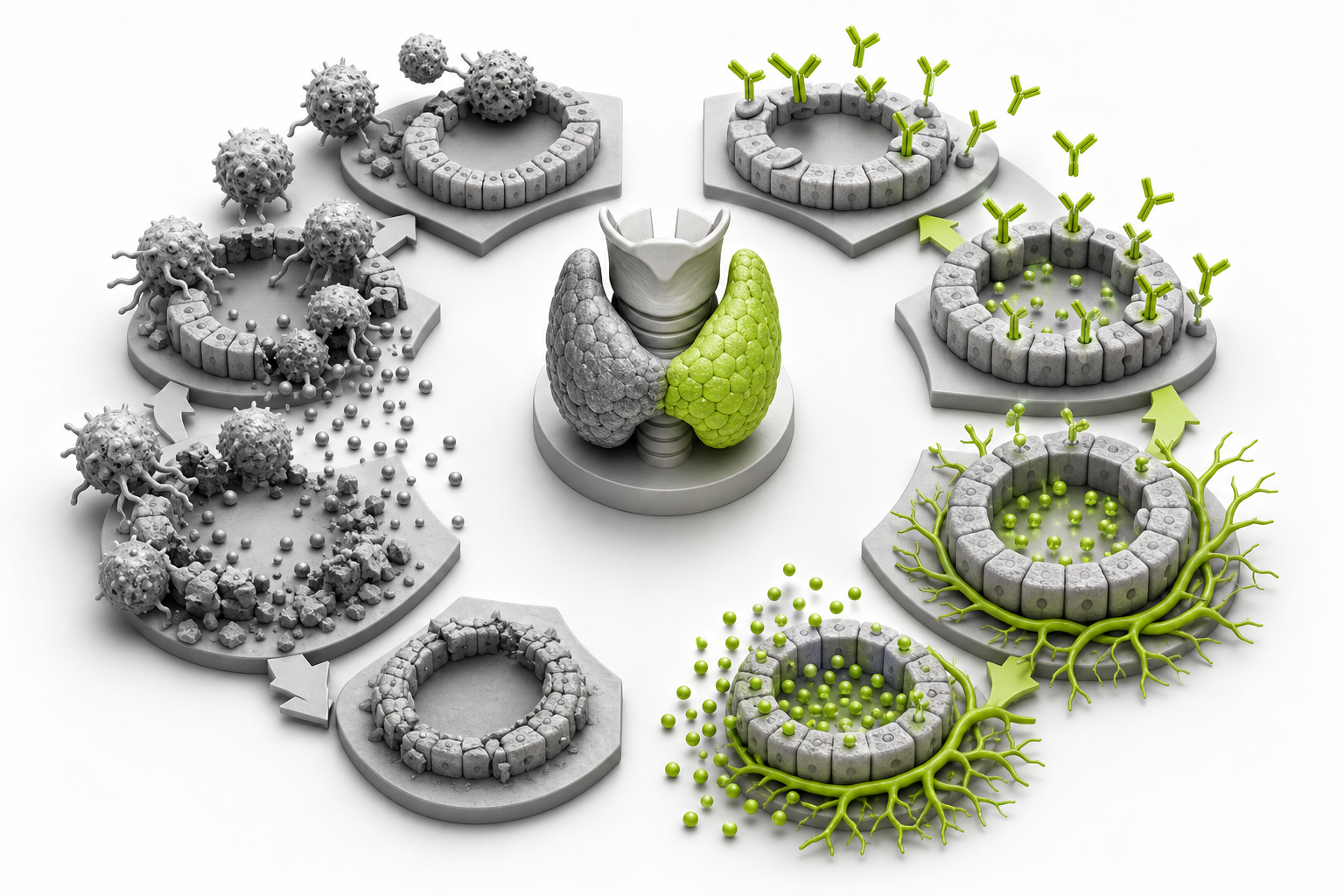
The fastest way to separate Hashimoto from Graves is to ask what the immune system is doing to the thyroid.
In **Hashimoto thyroiditis**, autoreactive lymphocytes progressively injure thyroid follicular cells. Anti–thyroid peroxidase antibodies (anti-TPO) and anti-thyroglobulin antibodies are characteristic markers of this process, but T-cell-mediated follicular destruction is central to the loss of functional thyroid tissue. The expected endpoint is primary hypothyroidism: the damaged gland produces too little T4, so the pituitary raises TSH in compensation. Histology classically shows lymphoid aggregates with germinal centers, follicular atrophy, and Hürthle cells—large follicular cells with granular eosinophilic cytoplasm caused by abundant mitochondria. These pathologic findings are summarized in a peer-reviewed review of Hashimoto thyroiditis.
In **Graves disease**, the immune system produces stimulating antibodies against the TSH receptor. These thyroid-stimulating immunoglobulins, or TSI, activate the receptor as though TSH were continuously present. The gland therefore grows, traps iodine, and synthesizes excess T3 and T4 despite suppression of pituitary TSH. The National Institute of Diabetes and Digestive and Kidney Diseases describes TSI as an antibody that mimics TSH and drives excessive thyroid hormone production.
A useful mental model is:
- **Hashimoto:** the immune system damages the hormone factory.
- **Graves:** the immune system holds the factory’s stimulation switch in the “on” position.
That distinction predicts the antibodies, thyroid-function tests, radioactive iodine uptake, and treatment.
Hashimoto vs Graves disease comparison
| Feature | Hashimoto thyroiditis | Graves disease | |---|---|---| | Core immune mechanism | Follicular destruction through cellular and humoral autoimmunity | TSH-receptor stimulation by functional autoantibodies | | Characteristic antibodies | Anti-TPO and anti-thyroglobulin | TSI; detectable as stimulating TSH-receptor antibodies within the broader TRAb category | | Typical TSH | Increased | Decreased | | Typical free T4 | Decreased in overt disease; may remain normal in subclinical disease | Increased | | Hyperthyroid phase | Possible but usually transient because stored hormone leaks from damaged follicles | Persistent unless treated because new hormone synthesis is increased | | Radioactive iodine uptake during thyrotoxicosis | Low in destructive thyroiditis | Diffusely increased | | Thyroid examination | Usually painless, firm, and diffusely enlarged early; may become atrophic | Painless diffuse goiter, often with a bruit or increased vascularity | | Distinctive extrathyroidal clues | Association with other autoimmune diseases | Graves orbitopathy and pretibial myxedema | | Classic histology | Lymphoid follicles with germinal centers and Hürthle cells | Tall crowded follicular epithelium with scalloped colloid | | Typical long-term treatment | Levothyroxine when hypothyroidism is present | Antithyroid medication, radioactive iodine, or thyroidectomy, depending on the clinical setting |
Antibody results must be interpreted with the physiology rather than used as isolated labels. Anti-TPO antibodies strongly support autoimmune thyroid disease, especially Hashimoto, but they may also occur in Graves. In contrast, a stimulating TSH-receptor antibody is the mechanistically important antibody in Graves. MedlinePlus explains the diagnostic roles of anti-TPO, anti-thyroglobulin, and TSH-receptor antibodies.
Read TSH and free T4 before naming the disease
For Step 1 reasoning, begin with the thyroid-function pattern and then determine its cause.
Primary hypothyroidism
The usual Hashimoto pattern is:
- **TSH increased**
- **Free T4 decreased**
A high TSH means the pituitary is appropriately trying to stimulate an underperforming thyroid. If free T4 is still normal, the patient has **subclinical hypothyroidism**. As follicular destruction progresses, free T4 falls and overt hypothyroidism develops.
Typical clinical findings include fatigue, cold intolerance, constipation, dry skin, weight gain, bradycardia, and delayed relaxation of deep tendon reflexes. The thyroid is generally non-tender. The NIDDK Hashimoto disease review notes that the gland may initially enlarge but can shrink after years of autoimmune damage.
Primary hyperthyroidism
The expected Graves pattern is:
- **TSH decreased**
- **Free T4 and/or T3 increased**
Excess circulating thyroid hormone suppresses pituitary TSH through negative feedback. Symptoms include heat intolerance, weight loss despite appetite, tremor, tachycardia, anxiety, sweating, and frequent bowel movements.
Diffuse goiter can occur in both diseases, so gland enlargement alone does not distinguish them. Graves-specific clues include proptosis or other features of orbitopathy, pretibial myxedema, a thyroid bruit, and markedly increased gland vascularity. Eye disease can exist even when thyroid hormone levels are normal, so it reflects autoimmune activity in orbital tissue rather than merely the systemic effects of excess T3 and T4.
Hashitoxicosis is release, not overproduction
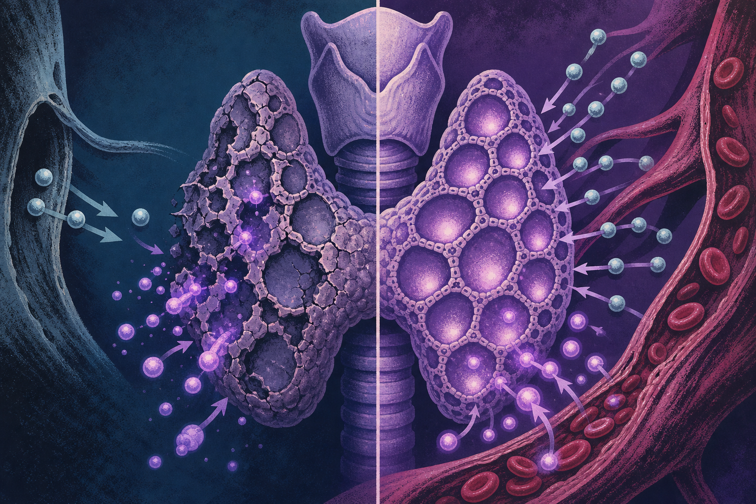
**Hashitoxicosis** is a transient thyrotoxic phase of Hashimoto thyroiditis. Immune-mediated injury disrupts follicles, allowing previously synthesized T3 and T4 to escape into the bloodstream. TSH falls because the pituitary senses the excess circulating hormone.
This can temporarily produce the same biochemical pattern seen in Graves:
- Low TSH
- High free T4 and sometimes high T3
- Palpitations, tremor, heat intolerance, or anxiety
The diseases diverge when the thyroid’s activity is measured. Graves involves active hormone synthesis and therefore produces **diffusely increased radioactive iodine uptake**. Destructive thyroiditis releases preformed hormone, so the injured gland has **low uptake**. Reviews of thyrotoxicosis describe this low-uptake pattern when excess hormone comes from follicular leakage rather than increased synthesis, while Graves produces diffuse uptake throughout the gland (hyperthyroidism review).
This distinction also explains why a thionamide such as methimazole is appropriate for Graves but generally does not correct destructive Hashitoxicosis. Thionamides inhibit new thyroid hormone synthesis; they cannot stop hormone that has already been synthesized from leaking out of damaged follicles. If transient adrenergic symptoms require treatment, a beta-blocker may provide symptomatic control while the stored hormone is cleared.
Doppler ultrasound can support the same reasoning. Graves usually has markedly increased diffuse blood flow because the gland is actively stimulated. Destructive thyroiditis generally has less flow, although early autoimmune inflammation may produce some overlap. When the distinction remains uncertain, the combination of TSH-receptor antibody testing and uptake imaging is more informative than anti-TPO positivity alone. A review comparing hyperthyroid-phase Hashimoto with Graves emphasizes the combined use of serology and imaging.
Clinical clues that should change the diagnosis
The following findings are particularly useful when both conditions remain plausible:
- **Proptosis, diplopia, or restrictive eye symptoms:** strongly favors Graves.
- **Pretibial myxedema:** favors Graves.
- **Thyroid bruit or striking Doppler vascularity:** favors Graves.
- **Hürthle cells with germinal centers:** favors Hashimoto.
- **Progression from transient thyrotoxicosis to hypothyroidism:** favors destructive thyroiditis.
- **Diffuse high radioactive iodine uptake:** favors Graves.
- **Low uptake during thyrotoxicosis:** favors Hashitoxicosis, painless thyroiditis, subacute thyroiditis, exogenous hormone use, or another non-synthetic cause.
- **Painful, tender thyroid with systemic inflammation:** suggests subacute granulomatous thyroiditis rather than either classic Hashimoto or Graves.
Do not equate any hyperthyroid laboratory result with Graves automatically. **Thyrotoxicosis** means tissues are exposed to excess thyroid hormone; **hyperthyroidism** specifically means the thyroid is actively producing too much hormone. Graves causes both. Hashitoxicosis causes thyrotoxicosis without sustained hyperthyroid synthesis.
Treatment follows the source of the hormone abnormality
Hashimoto treatment is directed at the hormone deficiency that results from tissue destruction. Patients who have developed hypothyroidism are treated with levothyroxine, synthetic T4. TSH is generally reassessed after a dose is started or changed because the hypothalamic-pituitary-thyroid axis needs time to equilibrate. The NIDDK recommends thyroid testing about six to eight weeks after initiating or adjusting levothyroxine. Antibody positivity by itself does not necessarily require thyroid hormone replacement if thyroid function remains normal.
Graves treatment must reduce active hormone synthesis or remove functioning thyroid tissue. Options include thionamides, radioactive iodine, and thyroidectomy; beta-blockers control adrenergic symptoms but do not reduce hormone synthesis. According to the NIDDK Graves treatment summary, methimazole is the antithyroid drug used most often, while radioactive iodine and surgery provide definitive treatment in selected patients.
The high-yield pharmacology link is direct: methimazole and propylthiouracil inhibit thyroid peroxidase, reducing organification and coupling. Propylthiouracil also decreases peripheral conversion of T4 to T3. Those actions matter when the gland is synthesizing hormone, as in Graves—not when damaged follicles are simply spilling stored hormone.
For additional endocrine mechanisms and pathology comparisons, explore the CoreStepPrep Core Concepts.
Worked vignette: when anti-TPO does not mean Hashitoxicosis
A 29-year-old woman presents with three months of weight loss, heat intolerance, palpitations, and gritty eyes. Her pulse is 112/min. Examination shows a symmetric non-tender goiter, mild bilateral proptosis, and a fine hand tremor. Laboratory studies show undetectable TSH, elevated free T4, and positive anti-TPO antibodies. Radioactive iodine uptake is diffusely increased.
**Most likely diagnosis: Graves disease.**
The decisive clues are proptosis and diffuse high iodine uptake. Together, they indicate TSH-receptor-driven hormone synthesis. Anti-TPO positivity confirms thyroid autoimmunity but does not prove Hashimoto thyroiditis; patients with Graves may also have anti-TPO antibodies.
**Why the nearest distractor, Hashitoxicosis, loses:** Hashitoxicosis can produce low TSH, high free T4, a painless goiter, and anti-TPO positivity. However, it results from follicular destruction and leakage of stored hormone. Radioactive iodine uptake should therefore be low rather than diffusely increased, and Graves-type orbitopathy would not be expected.
If the vignette instead described transient palpitations followed by cold intolerance and an increasing TSH, with low uptake during the thyrotoxic phase, Hashitoxicosis would become the better answer.
Final takeaways
- Hashimoto destroys follicles, whereas Graves stimulates the TSH receptor.
- Anti-TPO and anti-thyroglobulin support Hashimoto; stimulating TSH-receptor antibodies support Graves.
- Typical Hashimoto causes high TSH and low free T4; Graves causes low TSH and high thyroid hormone levels.
- Hashitoxicosis releases stored hormone and has low uptake, while Graves synthesizes new hormone and has diffuse high uptake.
- Proptosis, pretibial myxedema, a thyroid bruit, and marked vascularity point toward Graves.
Ready to apply the distinction? Try a free USMLE question at CoreStepPrep.
Sources and further reading
- pubmed.ncbi.nlm.nih.gov/32805423
- www.niddk.nih.gov/health-information/endocrine-diseases/graves-disease
- medlineplus.gov/lab-tests/thyroid-antibodies
- www.niddk.nih.gov/health-information/endocrine-diseases/hashimotos-disease
- pmc.ncbi.nlm.nih.gov/articles/5014602
- pmc.ncbi.nlm.nih.gov/articles/PMC10259585