Glomerulonephritis: Mechanisms and USMLE Clues
Distinguish immune-complex, anti-GBM, and ANCA glomerulonephritis using complement levels, biopsy staining patterns, and a worked USMLE vignette.
Glomerulonephritis becomes easier to distinguish when you connect the immune mechanism to the kidney biopsy: immune-complex disease usually produces granular staining, anti-glomerular basement membrane disease produces linear staining, and ANCA-associated disease is typically pauci-immune. Complement levels and clinical clues help narrow the differential, but they do not replace the biopsy pattern.
The central Step 1 reasoning problem is not simply recognizing blood in the urine. It is identifying **what is attacking the glomerulus** when several diseases produce the same nephritic findings.
Start with the shared injury, then identify the mechanism
Glomerular inflammation disrupts the filtration barrier. Red blood cells and protein enter the urine, while reduced filtration and sodium retention can contribute to rising creatinine, edema, and hypertension. Dysmorphic red blood cells and red blood cell casts support a glomerular source of bleeding; they do not identify a specific immune mechanism. The CDC description of poststreptococcal glomerulonephritis illustrates this shared nephritic presentation. (cdc.gov)
When glomerular capillary injury is severe, breaks in the filtration barrier allow coagulation proteins to enter Bowman space. Fibrin formation and activation of parietal epithelial cells contribute to a cellular **crescent**; inflammatory cells may also accumulate. A crescent signals severe injury, but its shape alone does not tell you whether the cause is an immune complex, an anti-GBM antibody, or vasculitis. Research on crescent formation explains why these different processes can converge on the same lesion. (pmc.ncbi.nlm.nih.gov)
Rapidly progressive glomerulonephritis describes a clinical syndrome of rapidly declining kidney function, often over weeks to months. Crescentic glomerulonephritis describes a biopsy pattern. The two are closely related, but neither term names the underlying disease.
Compare the three immunofluorescence patterns
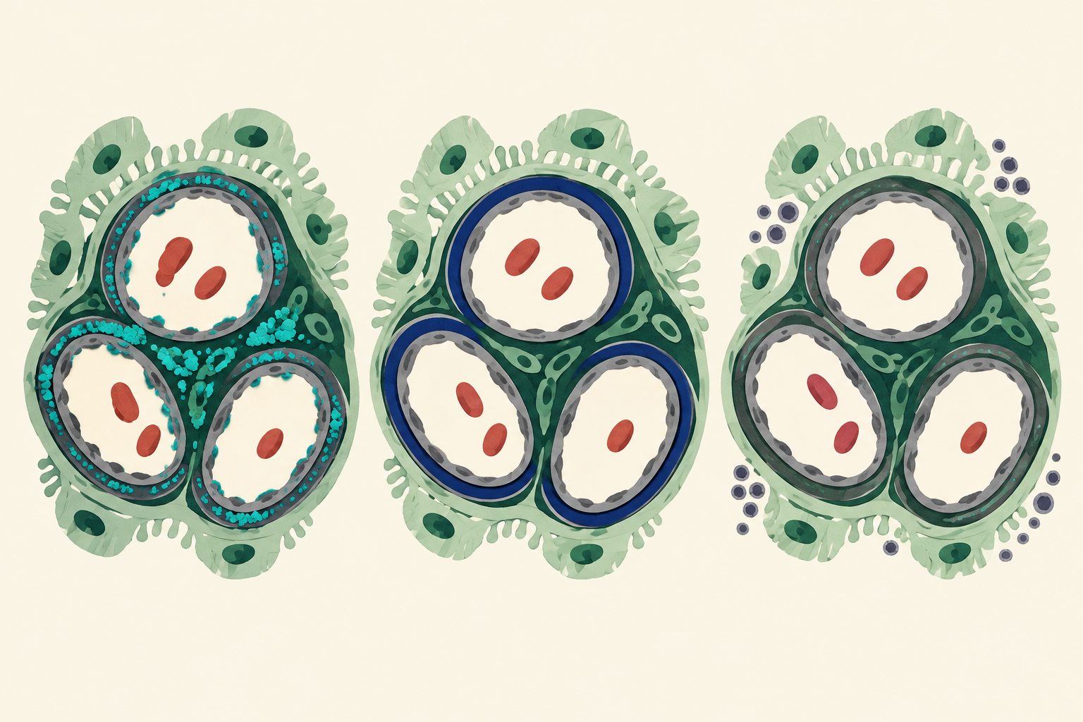
Immunofluorescence uses labeled antibodies to show where immunoglobulins and complement are present in a kidney specimen. The distribution offers a clue to the mechanism: patchy deposits, binding along the basement membrane, or little detectable deposited immunoglobulin. These are classic patterns, not absolute rules. The review of crescentic glomerulonephritis describes how biopsy distinguishes the major categories. (pubmed.ncbi.nlm.nih.gov)
| Mechanism | Typical immunofluorescence | Typical serum complement | Clues that support it | |---|---|---|---| | **Immune-complex mediated** | Granular immunoglobulin and/or complement deposits | Depends on the disease: often low in poststreptococcal GN and active lupus nephritis; usually normal in IgA nephropathy | Recent infection with a delay, mucosal-infection-associated hematuria, or systemic lupus features | | **Anti-GBM antibody mediated** | Smooth, linear IgG along the GBM | Usually normal | Rapidly progressive GN with circulating anti-GBM antibodies; pulmonary hemorrhage may occur | | **Pauci-immune, often ANCA associated** | Little or no glomerular immunoglobulin staining | Usually normal | Necrotizing GN with vasculitis findings, such as chronic sinus disease or pulmonary involvement |
Complement is a **supporting clue**, not a test that independently assigns a mechanism. Normal complement does not rule out immune-complex disease: IgA nephropathy is the important counterexample. Likewise, low serum complement can occur in other settings and should be interpreted alongside the clinical history, serology, and biopsy.
Why immune complexes produce granular staining
Immune-complex GN involves antibodies and antigens accumulating in or forming within glomerular tissue. Their deposits are discontinuous rather than evenly distributed along the entire basement membrane, giving the characteristic granular appearance. The location and contents of the deposits then help distinguish individual diseases.
Poststreptococcal GN: delayed hematuria and low C3
In the classic presentation, a child develops edema, dark urine, and hypertension after a symptom-free interval following a streptococcal infection. The interval matters: the CDC notes that poststreptococcal GN generally follows pharyngitis by about 10 days and can follow skin infection by up to 3 weeks. Serum C3 is typically low. (cdc.gov)
On biopsy, immunofluorescence classically shows coarse granular C3 with immunoglobulin, often IgG. Electron microscopy can reveal **subepithelial humps**: deposits on the urinary-space side of the GBM, beneath podocytes. A review of infection-related GN emphasizes that these findings are characteristic but not unique to poststreptococcal disease. (pmc.ncbi.nlm.nih.gov)
The reasoning is a chain rather than a single buzzword: a preceding infection, a delay, low C3, and granular deposits all support a postinfectious immune process. A positive streptococcal antibody result can support a recent infection; it is not the same thing as finding bacteria growing inside the glomerulus.
IgA nephropathy: hematuria close to a respiratory infection
IgA nephropathy is a useful distractor because it also can present with dark urine around a throat or respiratory illness. In the classic vignette, hematuria occurs **during the infection or within a few days**, rather than after the longer poststreptococcal interval. Episodes may recur, serum complement is usually normal, and biopsy shows dominant IgA deposits in the mesangium. The NIDDK guide to IgA nephropathy describes its association with respiratory infections and the role of biopsy in identifying deposits. (niddk.nih.gov)
Timing helps choose between the two, but it is not definitive in isolation. The stronger comparison combines timing with C3 and, when available, the deposit pattern: mesangial IgA versus the typical postinfectious C3-rich granular pattern and subepithelial humps.
Lupus nephritis: immune complexes with a systemic setting
Active lupus nephritis provides another contrast. A patient with systemic lupus erythematosus may have low C3 and C4, while biopsy can show a granular **full-house** immunofluorescence pattern: staining for IgG, IgA, IgM, C3, and C1q. Diffuse proliferative lupus nephritis can also show subendothelial deposits that create prominent capillary-wall “wire loops.” These findings reflect an immune-complex process, not direct anti-GBM antibody binding. (pubmed.ncbi.nlm.nih.gov)
Why anti-GBM staining is linear
In anti-GBM disease, the antibody target is an intrinsic component of the basement membrane, classically the alpha-3 chain of type IV collagen. Antibody binding follows the distribution of the target along the glomerular capillary wall, producing smooth **linear IgG** staining. This is different from the patchy appearance of immune-complex deposits. (pubmed.ncbi.nlm.nih.gov)
The disease can affect the kidneys, lungs, or both. Hemoptysis with rapidly worsening kidney function is therefore a strong clue, but absence of pulmonary hemorrhage does not exclude anti-GBM disease. Serum anti-GBM testing and kidney biopsy help establish the diagnosis; the NIDDK anti-GBM resource describes both the renal and pulmonary manifestations. (niddk.nih.gov)
Do not stop at “hematuria plus hemoptysis.” ANCA-associated vasculitis can also cause pulmonary-renal disease. The decisive distinction in a classic biopsy question is **linear IgG versus little or no immune staining**, interpreted with the antibody results.
Why ANCA-associated GN can be pauci-immune
In ANCA-associated vasculitis, circulating antibodies commonly recognize proteinase 3 (PR3) or myeloperoxidase (MPO). They participate in neutrophil activation and small-vessel inflammation, which can damage glomerular capillaries. Unlike immune-complex GN, the characteristic lesion has few or no immune deposits on immunofluorescence; this is what **pauci-immune** means. (pubmed.ncbi.nlm.nih.gov)
For example, granulomatosis with polyangiitis can combine chronic destructive sinonasal disease, lung abnormalities, and glomerulonephritis; PR3-ANCA is a classic association. Microscopic polyangiitis is often associated with MPO-ANCA. These associations help build a differential but are not exclusive pairings. (pmc.ncbi.nlm.nih.gov)
Two pitfalls follow from the mechanism. First, “pauci-immune” does **not** mean that the injury is nonimmune or that no antibody is present in the blood. It describes the scarcity of detectable glomerular immune deposits. Second, most—but not all—patients with pauci-immune crescentic GN have detectable ANCA. (pubmed.ncbi.nlm.nih.gov)
Worked vignette: pulmonary-renal disease with a decisive biopsy clue
A 37-year-old patient presents with fatigue, blood-streaked sputum, dark urine, and a rapidly rising creatinine level. Urinalysis shows red blood cell casts. Serum C3 and C4 are within the reference range. Kidney biopsy reveals crescents and smooth, continuous IgG staining along the glomerular capillary walls.
**Most likely mechanism:** antibodies directed against an intrinsic GBM antigen, as in anti-GBM disease.
**Decisive clue:** the *linear IgG* staining. Red blood cell casts establish glomerular bleeding, and crescents indicate severe injury, but neither identifies the cause. The continuous staining pattern points to antibody binding along the basement membrane.
**Why the nearest distractor loses:** ANCA-associated vasculitis can also cause hemoptysis, rapidly progressive GN, normal complement, and crescents. Its classic biopsy, however, is pauci-immune rather than strongly linear for IgG. Normal complement supports both possibilities, so it cannot settle the comparison.
**Why poststreptococcal GN is less likely:** its classic pattern combines a delayed presentation after streptococcal infection, low C3, and granular rather than linear staining. The vignette does not supply that sequence.
Why the distinction changes evaluation and treatment
Rapidly worsening kidney function with an active glomerular urine sediment warrants urgent evaluation. Clinicians combine kidney function and urine testing with targeted serologies—such as anti-GBM antibodies, ANCA, and complement—and often kidney biopsy. Results must be interpreted together: a normal complement level or a single antibody result should not be treated as a complete diagnosis. (niddk.nih.gov)
The mechanism also explains why treatments differ. Anti-GBM disease is often treated with plasma exchange to remove circulating antibodies, plus corticosteroids and cyclophosphamide to suppress the immune response. Severe ANCA-associated vasculitis is commonly treated with glucocorticoids and an immunosuppressive agent such as rituximab or cyclophosphamide; plasma exchange is not routinely added for every patient with ANCA-associated GN. Poststreptococcal GN management focuses on complications such as edema and hypertension, with antibiotics to eradicate remaining group A streptococci. See the NIDDK anti-GBM treatment summary, vasculitis management guideline, and CDC poststreptococcal guidance. (niddk.nih.gov)
For a broader renal foundation, explore the CoreStepPrep Core Concepts.
Key takeaways
- **Granular staining** suggests deposited immune material; use the clinical setting, complement, and deposit location to distinguish poststreptococcal GN, IgA nephropathy, and lupus nephritis.
- **Linear IgG** points toward antibodies binding the GBM in anti-GBM disease.
- **Pauci-immune staining** means few detectable glomerular immune deposits and commonly accompanies ANCA-associated vasculitis.
- **Crescents and red blood cell casts identify severe glomerular injury, not its specific cause.**
- Normal complement does not exclude immune-complex GN, and pulmonary-renal symptoms alone do not distinguish anti-GBM disease from ANCA-associated vasculitis.
Apply these distinctions to another clinical scenario: Try a free USMLE question at CoreStepPrep.
Sources and further reading
- www.cdc.gov/group-a-strep/hcp/clinical-guidance/post-streptococcal-glomerulonephritis.html
- pmc.ncbi.nlm.nih.gov/articles/PMC7170443
- pubmed.ncbi.nlm.nih.gov/22313839
- pmc.ncbi.nlm.nih.gov/articles/PMC9677723
- www.niddk.nih.gov/health-information/kidney-disease/iga-nephropathy
- www.niddk.nih.gov/health-information/kidney-disease/glomerular-disease/anti-gbm-goodpastures-disease
- pubmed.ncbi.nlm.nih.gov/26688808
- pmc.ncbi.nlm.nih.gov/articles/PMC12344527