G6PD Deficiency: Triggers, Bite Cells, and Clues
Learn how G6PD deficiency links oxidative triggers to Heinz bodies, bite cells, episodic hemolysis, and falsely normal enzyme tests on Step 1.
G6PD deficiency causes episodic hemolysis when oxidative stress overwhelms a red blood cell’s limited ability to regenerate reduced glutathione. On Step 1, connect the sequence **oxidant exposure → hemoglobin denaturation → Heinz bodies → splenic removal → bite cells**, and remember that G6PD activity may look normal during the acute episode because the most deficient cells have already been destroyed.
From oxidative stress to Heinz bodies and bite cells
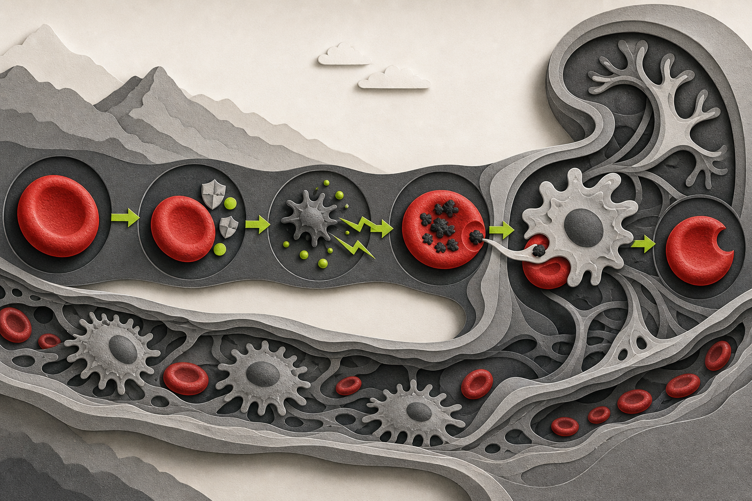
Glucose-6-phosphate dehydrogenase catalyzes the first and rate-limiting reaction of the oxidative phase of the pentose phosphate pathway:
**Glucose-6-phosphate + NADP⁺ → 6-phosphoglucono-δ-lactone + NADPH**
The important product for this disease is **NADPH**. Glutathione reductase uses NADPH to convert oxidized glutathione back to reduced glutathione, or GSH. Glutathione peroxidase can then use GSH to detoxify hydrogen peroxide and other reactive oxygen species.
Mature red blood cells rely heavily on this pathway for protection against oxidative injury. They continually carry oxygen, contain iron-rich hemoglobin, and lack the metabolic flexibility of nucleated cells. When G6PD activity is deficient, an oxidative challenge consumes reduced glutathione faster than it can be regenerated.
Oxidation then damages hemoglobin and the red cell membrane:
- Reactive oxygen species accumulate.
- Hemoglobin becomes oxidized and denatures.
- Denatured hemoglobin precipitates as **Heinz bodies**.
- Splenic macrophages remove Heinz body-containing portions of the membrane.
- The remaining red cell has a semicircular defect: a **bite cell**, also called a degmacyte.
This is why Heinz bodies and bite cells are related but not interchangeable. **Heinz bodies are the precipitated hemoglobin inside the cell; bite cells are the membrane shape left after the spleen removes that damaged material.** Heinz bodies generally require a supravital stain, whereas bite cells may be visible on a routine peripheral smear. A detailed review of oxidant injury and red-cell clearance describes how Heinz body formation reduces red-cell deformability and leads to macrophage-mediated removal.
Hemolysis may have both extravascular and intravascular components. Splenic clearance produces extravascular hemolysis and indirect hyperbilirubinemia, while severely injured cells may rupture in circulation and produce hemoglobinemia or dark, hemoglobin-containing urine.
The triggers that create oxidative stress
Most affected people are asymptomatic between episodes. Hemolysis appears when an infection, food, or medication sharply increases oxidative stress. The MedlinePlus Genetics description of G6PD deficiency identifies infections, certain medications, and fava beans as major precipitating categories.
Classic Step 1 associations include:
- **Infections:** Activated inflammatory cells generate oxidants, making bacterial or viral illness an important trigger even when no medication exposure is present.
- **Fava beans:** Favism results from oxidant compounds in fava beans and occurs only in susceptible individuals.
- **Antimalarials:** Primaquine is the classic association.
- **Antimicrobial drugs:** Dapsone, sulfonamide antibiotics, and nitrofurantoin are commonly tested examples.
- **Rasburicase:** Its metabolism of uric acid generates hydrogen peroxide, creating a dangerous oxidative challenge in G6PD-deficient red cells.
The unifying clue is not the specific drug name. It is a **new oxidative stressor followed by abrupt, nonimmune hemolysis**. A vignette may therefore use pneumonia or another infection without including a recognizable medication.
Symptoms reflect the sudden fall in red-cell mass and the products of hemoglobin breakdown. Patients may develop fatigue, pallor, jaundice, tachycardia, back or abdominal discomfort, and dark urine. Between episodes, the hemoglobin and peripheral smear may be normal.
The hemolysis pattern and smear findings
The laboratory pattern is that of hemolytic anemia:
- Decreased hemoglobin and hematocrit
- Increased reticulocyte count after the marrow responds
- Increased lactate dehydrogenase
- Increased indirect bilirubin
- Decreased haptoglobin, particularly with intravascular hemolysis
- Negative direct antiglobulin test because the process is not antibody-mediated
- Bite cells and sometimes blister cells on the routine smear
- Heinz bodies with a supravital stain
A negative direct antiglobulin test helps distinguish G6PD deficiency from warm autoimmune hemolytic anemia. It does not, by itself, establish G6PD deficiency; the oxidant exposure, episodic presentation, and characteristic smear provide the stronger combination.
| Disorder | Typical pattern | Smear or test clue | Distinction from G6PD deficiency | |---|---|---|---| | **G6PD deficiency** | Episodic hemolysis after oxidative stress | Bite cells; Heinz bodies with supravital stain; DAT negative | Triggered attacks with oxidant injury | | **Warm autoimmune hemolytic anemia** | Acquired extravascular hemolysis | Spherocytes; DAT positive for IgG, sometimes complement | Antibody-mediated rather than oxidant-mediated | | **Hereditary spherocytosis** | Chronic or intermittent extravascular hemolysis | Spherocytes, increased MCHC, DAT negative | Membrane defect; splenomegaly and family history may dominate | | **Microangiopathic hemolysis** | Intravascular fragmentation | Schistocytes, often thrombocytopenia | Mechanical shearing rather than hemoglobin denaturation | | **Pyruvate kinase deficiency** | Usually chronic congenital hemolysis | Echinocytes may appear; ATP deficiency | Glycolytic ATP failure rather than impaired NADPH production |
The highest-yield distinction is morphological: **oxidized hemoglobin produces Heinz bodies, and splenic removal of those inclusions produces bite cells**. Schistocytes instead indicate physical fragmentation, while spherocytes suggest membrane loss from hereditary or immune mechanisms.
Why G6PD testing can be falsely normal during hemolysis
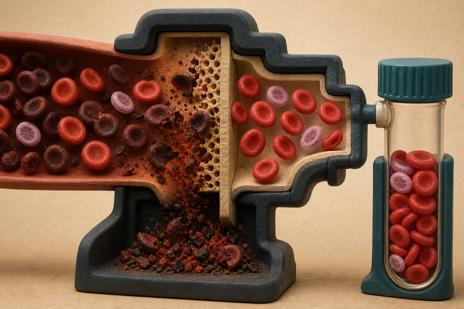
The enzyme assay has a classic timing pitfall. G6PD activity declines as red cells age, so older deficient erythrocytes are especially vulnerable to oxidant injury. During an acute episode, those cells are preferentially destroyed.
The blood sample obtained after hemolysis may therefore contain a disproportionate number of young erythrocytes and reticulocytes. These surviving cells have greater G6PD activity than the older cells that were removed. The measured average can fall within the normal range even though the patient has G6PD deficiency.
The practical interpretation is:
- **A low enzyme activity supports the diagnosis.**
- **A normal result during or soon after acute hemolysis does not reliably exclude it.**
- If clinical suspicion remains high, repeat quantitative testing after the hemolytic episode and reticulocytosis have resolved.
Official MedlinePlus guidance on G6PD testing specifically notes that testing may need to be repeated after an episode of hemolytic anemia. Recent transfusion can also obscure the result because donor red cells may have normal enzyme activity.
This pitfall is particularly important in a vignette that gives both a normal enzyme result and strong evidence of oxidant hemolysis. The normal result is not necessarily contradictory; it may be the clue that testing was performed too early.
X-linked inheritance and population associations
The G6PD gene is located on the X chromosome. Hemizygous males are more likely to have a clearly deficient red-cell population, and an affected father does not transmit the allele to his sons.
Heterozygous females have two red-cell populations because of random X-chromosome inactivation. Depending on lyonization, they may be asymptomatic, have intermediate enzyme activity, or experience clinically important hemolysis. Thus, being female does not exclude the diagnosis.
G6PD-deficiency variants are more prevalent in populations with ancestry from parts of Africa, the Mediterranean, the Middle East, and Asia. The geographic distribution reflects a selective advantage against severe malaria, although ancestry should function as a supporting clue rather than a diagnostic requirement. The MedlinePlus Genetics overview notes both the X-linked inheritance and the association between G6PD variants and partial malaria protection.
Management follows the trigger and severity
The first step in an acute episode is to remove the oxidant stressor when possible. That means stopping the responsible medication or treating the precipitating infection. Many attacks become self-limited as the vulnerable older cells are removed and younger red cells with greater enzyme activity predominate.
Supportive management may include hydration, monitoring hemoglobin and renal function, and transfusion for severe or symptomatic anemia. Neonatal hyperbilirubinemia requires bilirubin-directed treatment such as phototherapy or, in severe cases, exchange transfusion. The MedlinePlus clinical overview describes trigger avoidance, treatment of infection, discontinuation of offending medicines, and transfusion when necessary.
Long-term prevention centers on documenting the diagnosis and avoiding recognized triggers. Because medication risk may vary with dose, G6PD variant, and clinical circumstances, real-world prescribing decisions should use current drug-specific guidance rather than relying on a memorized list alone.
Worked vignette: the normal assay that does not rule it out
A 22-year-old man develops fatigue, jaundice, and dark urine three days after beginning nitrofurantoin for a urinary tract infection. Laboratory studies show decreased hemoglobin, reticulocytosis, increased indirect bilirubin and LDH, and decreased haptoglobin. The direct antiglobulin test is negative. A peripheral smear shows several erythrocytes with semicircular defects along their margins. A G6PD activity assay obtained during admission is reported as normal.
**Most likely diagnosis: G6PD deficiency with acute oxidant-induced hemolysis.**
The decisive clue is the combination of an oxidant exposure and **bite cells**. Nitrofurantoin supplied the oxidative challenge; denatured hemoglobin formed Heinz bodies; and splenic macrophages removed the inclusions, leaving bite-shaped membrane defects.
The normal enzyme assay does not eliminate the diagnosis because it was measured during active hemolysis. Older, highly deficient cells were preferentially destroyed, leaving reticulocytes and younger erythrocytes with relatively greater enzyme activity. The assay should be repeated after recovery if confirmation is still needed.
The nearest distractor is warm autoimmune hemolytic anemia. That disorder can also cause jaundice, reticulocytosis, low haptoglobin, and indirect hyperbilirubinemia. It loses because warm autoimmune hemolysis usually produces spherocytes and a positive direct antiglobulin test—not an oxidant-triggered episode with bite cells.
Final takeaways
- G6PD deficiency reduces NADPH production, impairing the regeneration of reduced glutathione.
- Oxidative stress denatures hemoglobin into Heinz bodies; splenic removal of those inclusions creates bite cells.
- Infections, fava beans, and oxidant medications can trigger abrupt, DAT-negative hemolysis.
- A normal G6PD assay during an acute episode may be falsely reassuring because young red cells and reticulocytes have relatively greater enzyme activity.
- Use exposure history, smear morphology, and the timing of testing together rather than relying on one isolated result.
For related pathway-based teaching, explore the CoreStepPrep Core Concepts.
Try a free USMLE question at CoreStepPrep