Knee Ligament Injuries: ACL, PCL, MCL, and LCL
Master ACL, PCL, MCL, and LCL knee injuries through mechanisms, exam maneuvers, imaging choices, management principles, and USMLE-style reasoning.
Knee ligament injuries become much easier to solve when you stop memorizing isolated test names and instead ask one biomechanical question: **Which abnormal tibial movement is the injured structure supposed to prevent?**
The ACL limits anterior tibial translation, the PCL limits posterior translation, the MCL resists valgus stress, and the LCL resists varus stress. From that framework, the mechanism, examination maneuver, and expected laxity follow logically. This approach is useful for both Step 1 anatomy questions and Step 2 CK diagnostic or management vignettes.
Match the Force to the Failed Restraint
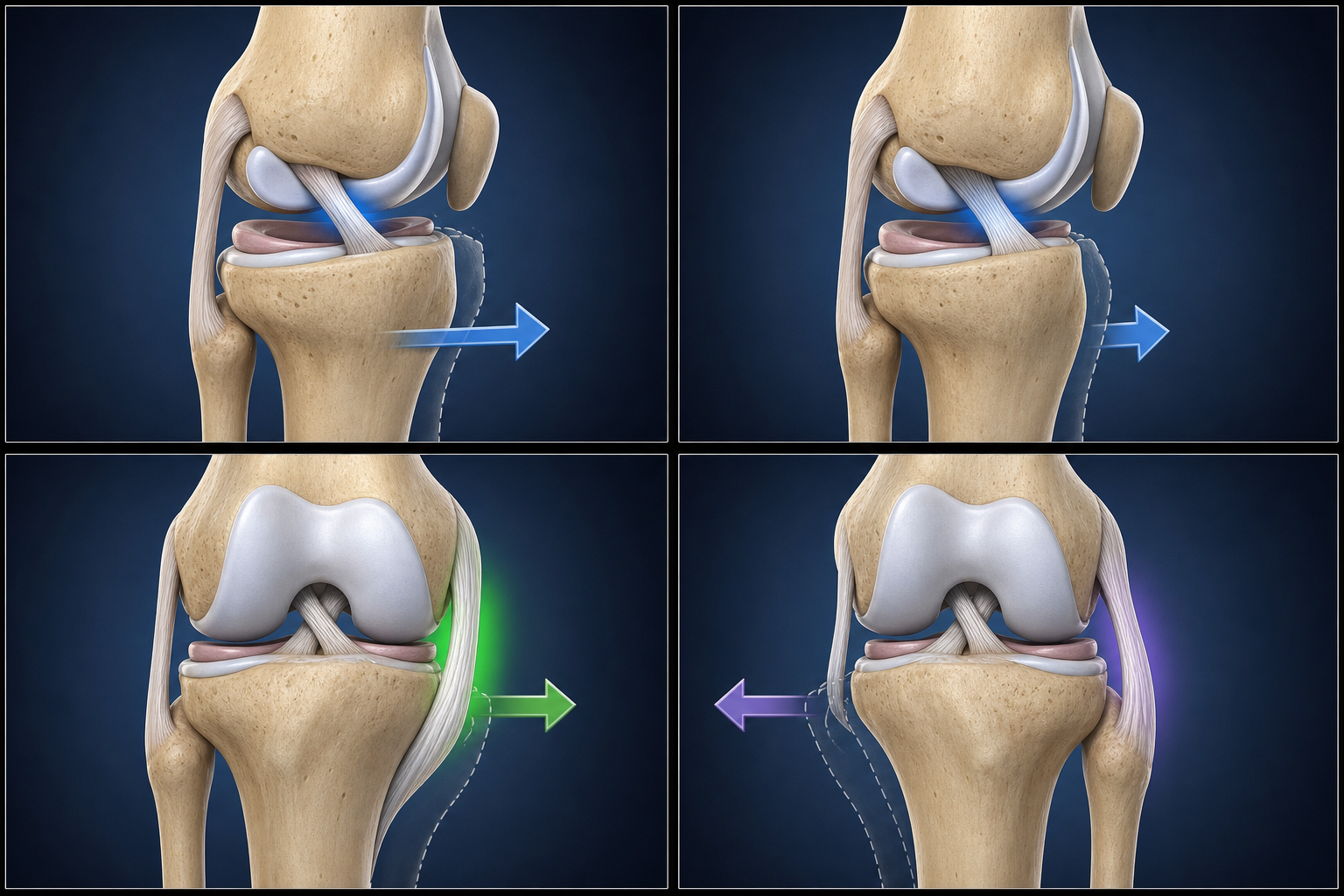
The cruciate ligaments primarily control anterior–posterior translation and contribute to rotational stability. The collateral ligaments stabilize the medial and lateral sides of the knee. These structures work together rather than in isolation, so high-energy injuries often damage more than one stabilizer. The core relationships are supported by established descriptions of knee ligament anatomy and biomechanics.
| Injured structure | Movement normally restrained | Typical mechanism | Key examination finding | |---|---|---|---| | ACL | Anterior translation of the tibia relative to the femur | Noncontact pivot, sudden deceleration, or landing with rotation | Positive Lachman; anterior drawer may also be positive | | PCL | Posterior translation of the tibia relative to the femur | Anterior force on a flexed tibia, such as a dashboard injury | Posterior sag and positive posterior drawer | | MCL | Valgus stress and medial joint opening | Blow to the lateral knee | Pain or laxity with valgus stress | | LCL | Varus stress and lateral joint opening | Blow to the medial knee | Pain or laxity with varus stress | | Meniscus | Load distribution and joint congruence rather than primary restraint of one directional force | Weight-bearing twist or deep flexion | Joint-line tenderness, catching, locking, or pain with provocative testing |
For exam questions, describe displacement from the perspective of the **tibia** unless the stem explicitly states otherwise. An ACL tear permits the tibia to move anteriorly; a PCL tear permits it to move posteriorly.
ACL Injury: Pivot, Hemarthrosis, and Anterior Laxity
The classic ACL mechanism is a noncontact change of direction with the foot planted. Sudden deceleration, awkward landing, and combined valgus–rotational loading are common versions of the same pattern. A patient may describe a pop followed by rapid swelling and difficulty continuing activity. Recurrent “giving way,” particularly during cutting movements, suggests functional instability.
Anatomically, the ACL runs from the anterior intercondylar region of the tibia to the posteromedial aspect of the lateral femoral condyle. Its disruption allows excessive anterior tibial translation and rotational instability.
Lachman, Anterior Drawer, and Pivot Shift Tests
The **Lachman test** is performed with the knee in approximately 20–30 degrees of flexion. The examiner stabilizes the distal femur and pulls the proximal tibia anteriorly. Increased translation or a soft endpoint supports ACL disruption.
The **anterior drawer test** evaluates anterior translation with the knee flexed to 90 degrees. It may be less useful immediately after injury because pain, swelling, or hamstring guarding can limit movement. Before interpreting the test, confirm that the tibia is not already sagging posteriorly from a PCL injury; pulling a posteriorly displaced tibia toward neutral can otherwise mimic an anterior drawer.
The **pivot shift test** assesses dynamic rotational instability. It is often difficult to perform in an acutely painful, guarded knee, but a clearly positive result strongly supports ACL deficiency. A systematic review of acute ACL examinations found that test performance varies: the pivot shift was relatively specific, while no single maneuver was perfect enough to replace the full history and examination. The reported findings reinforce using a combination of ACL examination maneuvers rather than relying on one result.
Imaging and ACL Management Decisions
Obtain radiographs when fracture, dislocation, or avulsion is possible. MRI is useful for directly evaluating the ACL and identifying associated meniscal, cartilage, collateral-ligament, or bone injuries. However, an ACL tear can often be recognized clinically; MRI should support the diagnostic and treatment plan rather than substitute for a mechanism-based examination.
Initial care generally focuses on controlling swelling, restoring range of motion, and recovering quadriceps activation. Long-term treatment is individualized. Persistent instability, participation in pivoting sports, occupational demands, associated repairable injuries, and patient goals may favor reconstruction, whereas selected patients can function well with structured rehabilitation. Current evidence does not support the blanket claim that every ACL rupture requires immediate surgery: a recent systematic review comparing reconstruction with rehabilitation alone found no clear difference in return-to-sport rates, although the certainty of evidence was low and treatment selection remains patient-specific.
PCL Injury: Recognize the Posterior Starting Position
The PCL is the primary restraint to posterior tibial translation. It originates from the medial femoral condyle and inserts on the posterior tibia.
A classic PCL injury occurs when an anterior force strikes the proximal tibia while the knee is flexed. The board-style example is a vehicle occupant whose flexed knee hits the dashboard. A fall directly onto a flexed knee can produce a similar force.
The patient may have posterior knee pain, swelling, or vague instability rather than the dramatic pivoting symptoms associated with ACL deficiency. Because isolated PCL injuries can be subtle, inspection is particularly important.
Posterior Sag and Posterior Drawer Tests
With the hips and knees flexed, view the knees from the side and compare the normal anterior prominence of the tibial plateau. In a PCL-deficient knee, gravity allows the tibia to settle posteriorly, producing the **posterior sag sign**.
For the **posterior drawer test**, flex the knee to 90 degrees and push the proximal tibia posteriorly. Excessive posterior translation supports PCL injury. Always determine the tibia’s resting position before applying force. If the tibia begins in a posteriorly subluxed position, the examination can be misread.
MRI can demonstrate an acute PCL tear and associated injuries, while radiographs help identify fractures or avulsions. Contemporary reviews emphasize that PCL assessment should combine the mechanism, posterior drawer or sag findings, comparison with the opposite knee, and appropriate imaging rather than depending on one test alone. Isolated lower-grade injuries are frequently treated nonoperatively, whereas marked instability and combined ligament injuries are more likely to require operative evaluation, as summarized in a current review of PCL assessment and management.
MCL and LCL Injuries: Valgus Versus Varus
Collateral-ligament questions usually test whether you can translate the direction of an external force into the side of the knee that opens.
A blow to the **lateral** knee drives the joint into valgus and stresses the **MCL**. A blow to the **medial** knee drives the joint into varus and stresses the **LCL**.
The MCL is broad and attached to the medial capsule; its deep fibers are related to the medial meniscus. The LCL, also called the fibular collateral ligament, runs from the lateral femoral epicondyle to the fibular head and is not attached to the lateral meniscus.
Stress Testing at 30 Degrees and Full Extension
Perform valgus and varus stress tests first with the knee flexed approximately 30 degrees. This position helps assess the collateral structures while reducing the contribution of other stabilizers.
- **Valgus stress:** medial pain or excessive medial joint opening suggests MCL injury.
- **Varus stress:** lateral pain or excessive lateral joint opening suggests LCL or posterolateral injury.
Repeat the maneuver near full extension when a more extensive injury is suspected. Laxity only in flexion is more compatible with an isolated collateral injury. Instability that persists in full extension should raise concern for additional capsular or cruciate damage.
Many isolated grade I and II MCL injuries heal with bracing as needed, early motion, and progressive rehabilitation. A systematic review of nonoperative MCL rehabilitation found substantial variation among protocols but confirmed that conservative treatment is widely used for isolated injuries.
An apparent LCL injury deserves additional attention because lateral instability may represent damage to the broader posterolateral corner. Examine for increased external rotation, varus thrust, peroneal nerve findings, and associated cruciate injury. MRI and stress radiographs can help characterize complex instability. A contemporary international posterolateral-corner consensus statement supports using multiple physical and radiographic assessments and individualized treatment for these injuries.
Meniscal Injury Versus Ligamentous Instability
A meniscal tear is favored by a weight-bearing twisting mechanism, joint-line pain, catching, or true mechanical locking. Swelling may develop more gradually than the rapid hemarthrosis associated with a major ACL injury, although timing alone cannot establish the diagnosis.
McMurray, Apley, and joint-line tenderness findings can support the diagnosis, but none should be treated as definitive in isolation. A systematic review of meniscal examination tests found variable accuracy and substantial heterogeneity across studies. On an exam, the combination of mechanism, joint-line symptoms, mechanical complaints, and provocative findings is more persuasive than one named maneuver.
MRI is the principal noninvasive study when the diagnosis remains uncertain, symptoms persist, or surgery is being considered. It is highly useful but not infallible; an updated meta-analysis of MRI for meniscal tears found high overall performance with differences between medial and lateral tears. Arthroscopy directly visualizes the meniscus and allows treatment, but it is an invasive procedure reserved for appropriately selected patients rather than a routine first diagnostic step.
The classic “unhappy triad” is ACL, MCL, and medial meniscus injury after valgus–rotational trauma. Preserve that association for board questions, but recognize that real multiligament injuries are more variable and may involve the lateral meniscus or anterolateral structures. One contemporary multiligament cohort found that lateral meniscal lesions were common in ACL plus medial-side injuries.
A Practical Examination Sequence
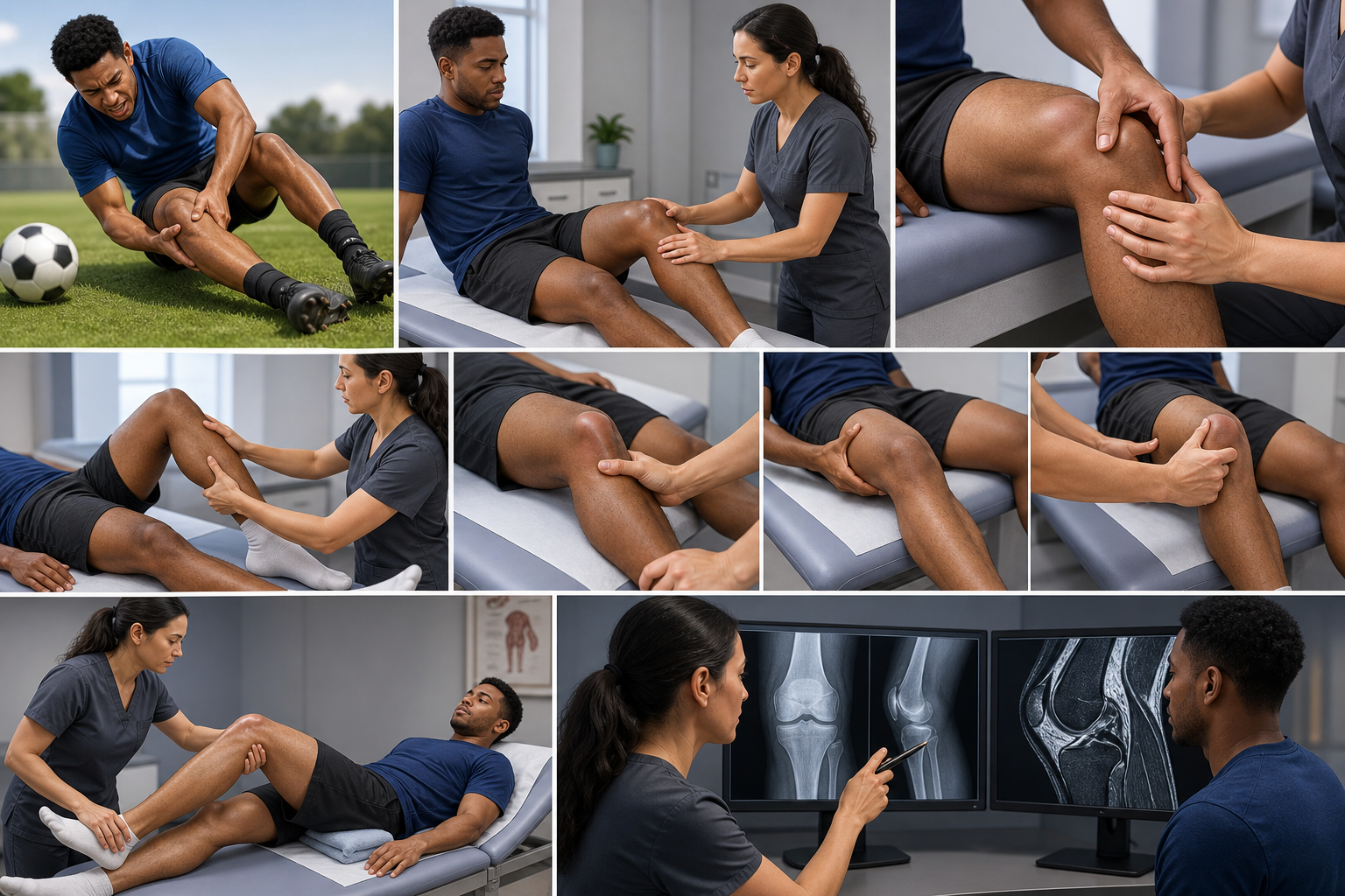
When a vignette gives you an injured, swollen knee, use the same sequence every time:
- **Identify the force:** pivot, anterior blow to a flexed tibia, lateral blow, medial blow, or weight-bearing twist.
- **Inspect the resting position:** look for swelling, deformity, bruising, and posterior sag.
- **Palpate deliberately:** check joint lines, ligament courses, fibular head, patella, and bony landmarks.
- **Test the predicted restraint:** Lachman, posterior drawer, valgus stress, or varus stress.
- **Look for associated injury:** meniscal symptoms, multiligament laxity, fracture, neurovascular deficits, or mechanical locking.
- **Choose imaging for the clinical question:** radiographs for bone and alignment; MRI for soft-tissue characterization and operative planning.
Exam-Style Vignette
A 21-year-old collegiate soccer player plants her right foot and turns sharply to follow the ball. She feels a pop and cannot continue playing. Within 90 minutes, the knee becomes markedly swollen. Examination shows increased anterior translation of the tibia with a soft endpoint when the knee is flexed 25 degrees. Valgus and varus stress tests show no laxity.
**Most likely injury:** ACL tear.
**Reasoning:** A noncontact planted-foot pivot followed by rapid swelling and instability strongly suggests ACL disruption. The examination maneuver described is the Lachman test, and increased anterior tibial translation with a soft endpoint is the key finding. Normal collateral stress tests make an isolated MCL or LCL tear less likely. MRI may define associated injuries, but the mechanism and examination already establish a strong clinical diagnosis.
Final Takeaways
- Translate each mechanism into the abnormal tibial movement it produces.
- ACL injury causes anterior laxity; PCL injury causes posterior sag and posterior laxity.
- Valgus stress tests the MCL, while varus stress tests the LCL and lateral stabilizers.
- Use physical tests as parts of a coherent history and examination, not as isolated diagnostic shortcuts.
- Management depends on instability, associated injuries, activity demands, and patient goals—not merely the ligament’s name.
Build your next study block with CoreStepPrep.