Demyelinating and Peripheral Neuropathies | USMLE
Distinguish MS, Guillain-Barré syndrome, myasthenia gravis, and CMT using localization, clinical patterns, diagnostic tests, and treatment clues.
Demyelinating and peripheral neuropathies become much easier when you stop memorizing isolated disease lists and first localize the lesion. Is the problem in the central nervous system, the peripheral nerve, or the neuromuscular junction? That single decision separates multiple sclerosis (MS), Guillain–Barré syndrome (GBS), Charcot–Marie–Tooth disease (CMT), and myasthenia gravis (MG) before MRI, cerebrospinal fluid, or antibody results appear.
For USMLE questions, build the diagnosis from four variables: **anatomic location, time course, examination pattern, and confirmatory test**. Then match treatment to the mechanism—acute inflammation, chronic autoimmune activity, impaired neuromuscular transmission, or inherited nerve dysfunction.
Localize Before You Name the Disease
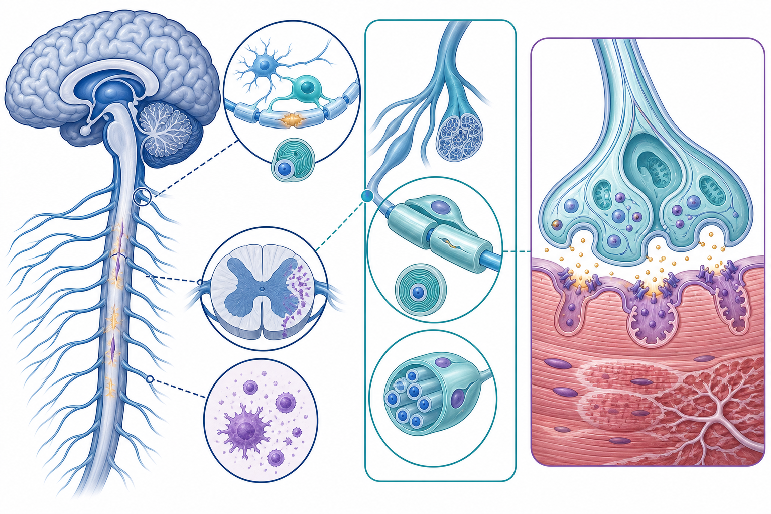
Myelin performs the same broad function throughout the nervous system—accelerating saltatory conduction—but different cells produce it in different locations:
- **Oligodendrocytes** myelinate axons in the CNS. Immune-mediated CNS demyelination points toward MS.
- **Schwann cells** myelinate peripheral nerves. Acute inflammatory demyelination in the PNS points toward GBS.
- **The neuromuscular junction** contains no myelin. MG is included because it produces weakness that can resemble peripheral nerve disease, but its pathology is postsynaptic.
- **Inherited peripheral nerve defects** can disrupt myelin or axons over years, as in CMT.
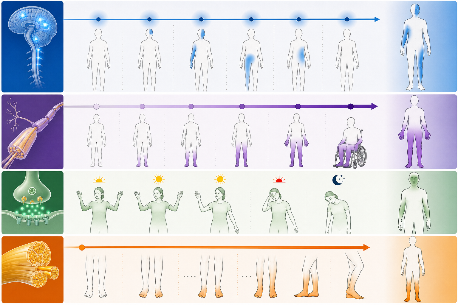
This localization predicts the examination. CNS disease can produce upper motor neuron signs, sensory levels, visual symptoms, or long-tract deficits. Peripheral neuropathy more often causes reduced reflexes, distal sensory loss, and lower motor neuron weakness. Neuromuscular-junction disease causes fatigable weakness without objective sensory loss.
Time course adds another layer. MS typically produces neurologic episodes separated in time and anatomic location. GBS progresses over days to several weeks. MG fluctuates during the day or with repeated muscle use. CMT evolves slowly over years.
Multiple Sclerosis Produces Multifocal CNS Deficits
MS is an immune-mediated disease in which inflammation, demyelination, and axonal injury occur within the brain, optic nerves, and spinal cord. The high-yield pathologic contrast is **oligodendrocyte injury in MS versus Schwann-cell and peripheral nerve involvement in GBS**.
A classic vignette describes a young adult with neurologic deficits that cannot be explained by one CNS lesion. Common presentations include:
- Painful monocular vision loss from optic neuritis
- Internuclear ophthalmoplegia from a medial longitudinal fasciculus lesion
- Limb weakness, spasticity, or hyperreflexia
- Focal sensory symptoms
- Ataxia or intention tremor
- Urinary urgency, retention, or other bladder dysfunction
- Worsening of prior deficits with heat, called the Uhthoff phenomenon
The core diagnostic concept is evidence of characteristic CNS injury with no better alternative explanation. MRI demonstrates lesions in typical locations, classically including periventricular, cortical or juxtacortical, infratentorial, and spinal cord regions. Contemporary diagnostic criteria incorporate clinical findings, MRI distribution, and CSF biomarkers; the current framework has expanded beyond the older board shorthand, but dissemination across characteristic CNS locations remains central to exam reasoning. The 2024 McDonald criteria discussion describes newer roles for optic nerve involvement, advanced imaging markers, oligoclonal bands, and kappa free light chains.
On standard USMLE-style questions, look for **ovoid periventricular plaques**, lesions of different ages, or simultaneous enhancing and nonenhancing lesions. CSF-restricted oligoclonal IgG bands support intrathecal immunoglobulin production, but they are supportive rather than uniquely specific to MS.
Acute Relapse and Long-Term Disease Control Are Different Decisions
A clinically meaningful acute relapse is treated with high-dose corticosteroids, commonly methylprednisolone, after excluding infection or another cause of transient worsening. A pseudo-relapse—reappearance of old symptoms during fever, heat exposure, or metabolic stress—does not necessarily represent new inflammatory injury. Recommendations for evaluating and treating relapses emphasize this distinction and the role of high-dose corticosteroid therapy in appropriate attacks (MS relapse recommendations).
Disease-modifying therapy is a separate long-term decision intended to reduce inflammatory disease activity. Agents encountered on exams include interferon beta, glatiramer acetate, natalizumab, and ocrelizumab, although real-world selection depends on disease phenotype, activity, comorbidities, pregnancy considerations, monitoring requirements, and adverse-effect profiles.
Natalizumab is especially testable because blocking leukocyte migration into the CNS can increase the risk of progressive multifocal leukoencephalopathy. The FDA’s natalizumab safety summary identifies anti-JC-virus antibodies, longer treatment duration, and prior immunosuppressant exposure as important PML risk factors.
**Exam pitfall:** Corticosteroids treat an acute MS relapse but are not a substitute for long-term disease-modifying therapy.
Guillain–Barré Syndrome Is an Acute Peripheral Nerve Emergency
GBS is an acute immune-mediated polyradiculoneuropathy, often occurring after a gastrointestinal or respiratory infection. Molecular mimicry can generate antibodies that cross-react with peripheral nerve components. The classic demyelinating form is acute inflammatory demyelinating polyradiculoneuropathy, although axonal variants also occur.
The typical pattern is:
- Symmetric weakness beginning in the legs
- Progression upward over days
- Decreased or absent deep-tendon reflexes
- Mild paresthesias or sensory symptoms despite prominent weakness
- Possible facial, bulbar, respiratory, or autonomic involvement
Unlike MS, GBS is a PNS process and therefore does not produce an upper motor neuron pattern. Areflexia is one of the strongest localization clues.
CSF classically shows **albuminocytologic dissociation**: elevated protein with few or no white blood cells. Protein may still be normal early in the illness, so an initially unrevealing lumbar puncture does not exclude GBS. Electrodiagnostic testing can support the diagnosis by demonstrating demyelinating or axonal peripheral nerve abnormalities.
The immediate danger is not the leg weakness itself. Respiratory muscle failure, impaired airway protection, arrhythmias, and extreme blood-pressure fluctuations can develop rapidly. Serial respiratory assessment—including bedside spirometry when feasible—should accompany repeated evaluation of bulbar function and autonomic stability.
The European Academy of Neurology and Peripheral Nerve Society guideline supports IV immunoglobulin or plasma exchange for patients with clinically significant weakness and recommends against oral corticosteroids. IVIG and plasma exchange are alternatives; routinely giving both sequentially does not provide an established advantage.
**Exam pitfall:** Do not wait for respiratory distress before assessing respiratory function. Also remember that corticosteroids help MS relapses but do not improve typical GBS outcomes.
Myasthenia Gravis Mimics Neuropathy Without Sensory Loss
MG is not a demyelinating disorder. It is an autoimmune neuromuscular-junction disease, most often caused by antibodies against postsynaptic acetylcholine receptors. Some patients instead have antibodies to muscle-specific kinase or other postsynaptic proteins.
The defining pattern is **fluctuating, fatigable weakness**:
- Ptosis or diplopia that worsens with sustained gaze
- Difficulty chewing near the end of a meal
- Dysarthria or dysphagia after repeated use
- Proximal limb weakness
- Improvement after rest
- Preserved sensation
- Usually preserved reflexes
Serum acetylcholine-receptor antibodies are the usual initial confirmatory study in generalized disease. Repetitive nerve stimulation may show a decremental response, while single-fiber electromyography is highly sensitive. Edrophonium is historically famous but is now rarely used; modern diagnosis relies more heavily on the clinical pattern, antibodies, and electrodiagnostic testing, as reviewed in current diagnostic approaches to MG.
Chest imaging is performed to evaluate the thymus because MG is associated with thymic hyperplasia and thymoma. Pyridostigmine improves neuromuscular transmission, while corticosteroids and steroid-sparing immunotherapies address autoimmune activity. Thymoma generally requires thymectomy. Thymectomy should also be discussed in selected patients with generalized acetylcholine-receptor-antibody-positive MG; an American Academy of Neurology practice advisory found benefit when thymectomy was added to prednisone in the studied nonthymomatous population.
Myasthenic crisis is severe respiratory or bulbar weakness requiring urgent airway assessment and rapid immunomodulation with IVIG or plasma exchange. Current myasthenic syndrome guidance also emphasizes that treatment selection depends on disease activity, antibody status, and thymic pathology.
The classic comparison is Lambert–Eaton myasthenic syndrome. MG causes postsynaptic dysfunction, weakness that worsens with use, and generally preserved reflexes. Lambert–Eaton involves presynaptic voltage-gated calcium channels, often causes autonomic symptoms and reduced reflexes, and may transiently improve with repeated activation.
Charcot–Marie–Tooth Disease Follows a Chronic Inherited Pattern
CMT describes a genetically heterogeneous group of hereditary motor and sensory neuropathies. CMT1A, a common demyelinating subtype, results from duplication involving **PMP22**, causing abnormal PMP22 dosage and impaired Schwann-cell myelination (PMP22 duplication research).
The phenotype is determined by length-dependent peripheral nerve dysfunction:
- Slowly progressive distal leg weakness
- Foot drop and frequent tripping
- Calf atrophy producing a “stork-leg” appearance
- Pes cavus and hammertoes
- Distal sensory loss
- Reduced ankle reflexes
- Later hand weakness in some patients
- Similar findings in relatives
The chronic course is the key discriminator. GBS develops over days to weeks; CMT progresses over years and is often accompanied by longstanding foot deformities.
Evaluation combines the phenotype, family history, nerve-conduction studies, and genetic testing. Current CMT clinical practice guidance recommends a sequential genetic approach and emphasizes individualized rehabilitation, exercise, orthoses, and orthopedic management. No broadly applicable curative pharmacologic treatment is currently established.
A High-Yield Differential for Weakness and Demyelination
| Disorder | Localization | Typical time course | Reflexes | Sensory findings | Key test clue | Core treatment principle | |---|---|---|---|---|---|---| | Multiple sclerosis | CNS myelin | Relapses or progression | Often increased in affected limbs | CNS sensory deficits possible | Characteristic MRI lesions; CSF-restricted oligoclonal bands | Steroids for relapse; disease-modifying therapy for long-term control | | Guillain–Barré syndrome | Nerve roots and peripheral nerves | Days to 4 weeks | Reduced or absent | Mild paresthesias common | CSF high protein with few cells; electrodiagnostic abnormalities | Respiratory monitoring plus IVIG or plasma exchange | | CIDP | Peripheral nerves | More than 8 weeks or relapsing | Reduced or absent | Often present | Demyelination on nerve-conduction studies | Chronic immunomodulatory therapy | | Myasthenia gravis | Postsynaptic neuromuscular junction | Fluctuating, use-dependent | Usually normal | Absent | AChR or MuSK antibodies; decrement on repetitive stimulation | Pyridostigmine and immune-directed therapy | | Lambert–Eaton syndrome | Presynaptic neuromuscular junction | Subacute or chronic | Reduced; may facilitate | No primary sensory loss | Facilitation with rapid stimulation | Search for malignancy; improve transmission and treat cause | | Charcot–Marie–Tooth disease | Inherited peripheral nerve disease | Years | Reduced distally | Distal sensory loss | Family history, nerve studies, genetic testing | Rehabilitation, orthoses, and genetic counseling |
A useful timing rule is that weakness progressing beyond eight weeks should prompt reconsideration of GBS and evaluation for acute-onset chronic inflammatory demyelinating polyradiculoneuropathy or another diagnosis.
Exam-Style Vignette and Reasoning
A 29-year-old man develops tingling in both feet nine days after recovering from diarrhea. Over three days, he develops difficulty climbing stairs and then raising his arms. Examination shows symmetric leg-predominant weakness and absent patellar and Achilles reflexes. Sensation is mildly reduced in the toes. He becomes intermittently tachycardic, and his blood pressure fluctuates during observation.
**What is the best next management approach?**
This is GBS: a recent gastrointestinal illness is followed by rapidly ascending symmetric weakness, areflexia, mild sensory symptoms, and autonomic instability. The next priorities are serial respiratory and bulbar assessment, close cardiac and blood-pressure monitoring, and prompt treatment with IVIG or plasma exchange when indicated by disease severity. Corticosteroids should not be selected.
Do not be distracted by the absence of reported CSF results. The bedside pattern already localizes the lesion to peripheral nerves, and urgent monitoring should not be delayed for lumbar puncture.
Final Takeaways
- First distinguish **CNS, peripheral nerve, and neuromuscular-junction** disease.
- MS produces multifocal CNS findings; GBS produces acute ascending weakness with areflexia.
- MG causes fatigable weakness without sensory loss, while CMT causes chronic distal weakness, sensory loss, and foot deformities.
- Match therapy to the time course: steroids for an MS relapse, IVIG or plasma exchange for GBS or myasthenic crisis, and rehabilitation for CMT.
- In rapidly progressive weakness, respiratory and bulbar assessment comes before diagnostic perfection.
Build your next study block with CoreStepPrep.
Sources and further reading
- pubmed.ncbi.nlm.nih.gov/42178152
- pubmed.ncbi.nlm.nih.gov/35055321
- www.fda.gov/news-events/press-announcements/fda-approves-first-biosimilar-treat-multiple-sclerosis
- pubmed.ncbi.nlm.nih.gov/37814552
- pubmed.ncbi.nlm.nih.gov/29655449
- pubmed.ncbi.nlm.nih.gov/32213645
- pubmed.ncbi.nlm.nih.gov/38152089
- pubmed.ncbi.nlm.nih.gov/38743588
- pubmed.ncbi.nlm.nih.gov/38431252